. 2017 Sep 19;8(1):598.
doi: 10.1038/s41467-017-00590-9.

Cross-talk between miR-471-5p and autophagy component proteins regulates LC3-associated phagocytosis (LAP) of apoptotic germ cells

Affiliations

Cross-talk between miR-471-5p and autophagy component proteins regulates LC3-associated phagocytosis (LAP) of apoptotic germ cells

Subbarayalu Panneerdoss et al. Nat Commun. .

Abstract

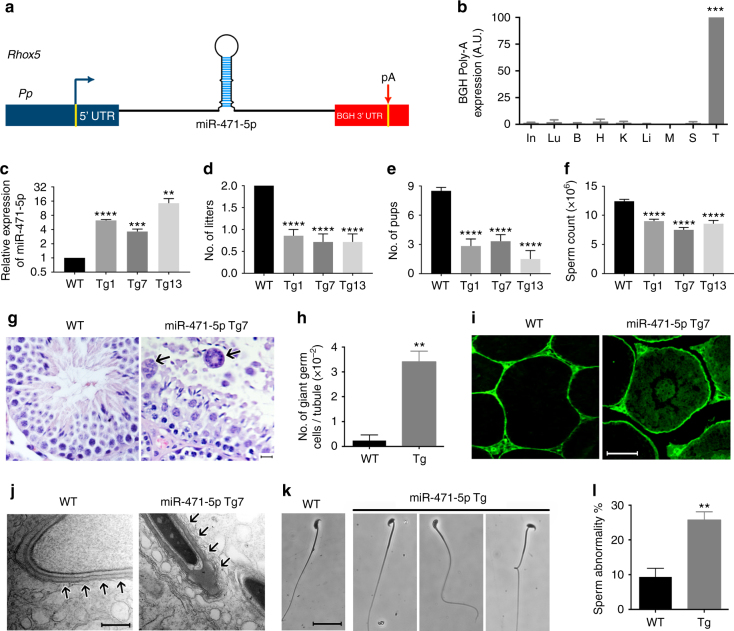
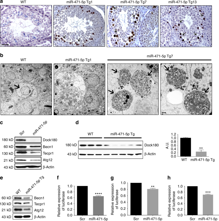
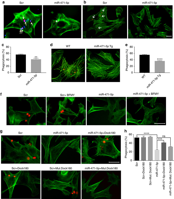
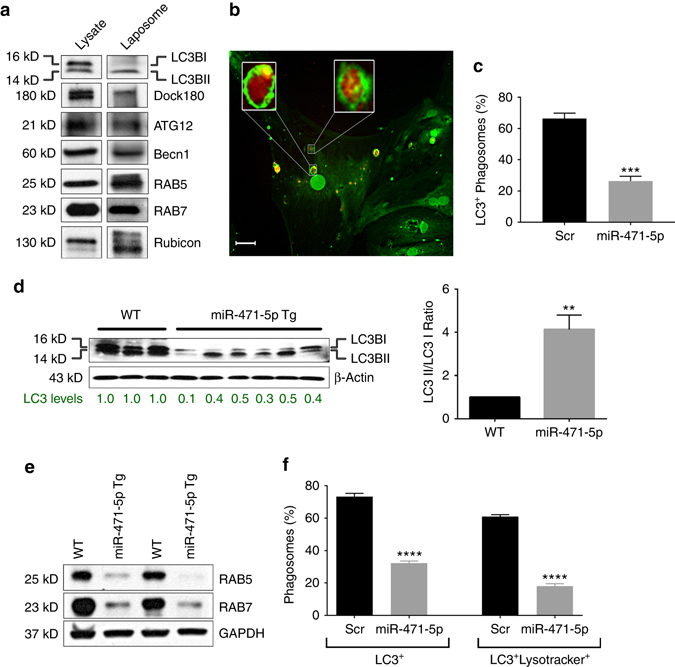
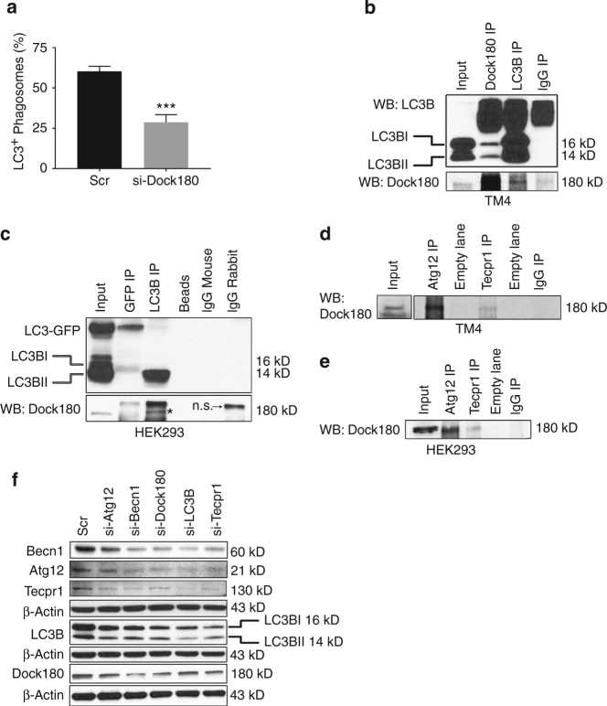
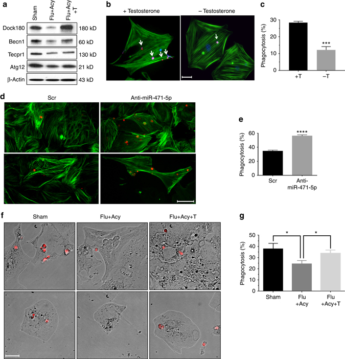
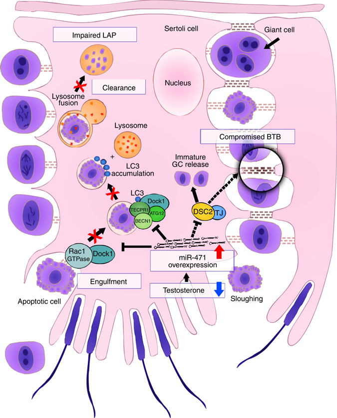
Phagocytic clearance of apoptotic germ cells by Sertoli cells is vital for germ cell development and differentiation. Here, using a tissue-specific miRNA transgenic mouse model, we show that interaction between miR-471-5p and autophagy member proteins regulates clearance of apoptotic germ cells via LC3-associated phagocytosis (LAP). Transgenic mice expressing miR-471-5p in Sertoli cells show increased germ cell apoptosis and compromised male fertility. Those effects are due to defective engulfment and impaired LAP-mediated clearance of apoptotic germ cells as miR-471-5p transgenic mice show lower levels of Dock180, LC3, Atg12, Becn1, Rab5 and Rubicon in Sertoli cells. Our results reveal that Dock180 interacts with autophagy member proteins to constitute a functional LC3-dependent phagocytic complex. We find that androgen regulates Sertoli cell phagocytosis by controlling expression of miR-471-5p and its target proteins. These findings suggest that recruitment of autophagy machinery is essential for efficient clearance of apoptotic germ cells by Sertoli cells using LAP.Although phagocytic clearance of apoptotic germ cells by Sertoli cells is essential for spermatogenesis, little of the mechanism is known. Here the authors show that Sertoli cells employ LC3-associated phagocytosis (LAP) by recruiting autophagy member proteins to clear apoptotic germ cells.


Conflict of interest statement

The authors declare no competing financial interests.

Figures

Fig. 1
Fig. 1
Reproductive defects in miR-471-5p transgenic (Tg) mice. a Schematic of the construct used to generate miR-471-5p transgenic mice (miR-471-5pTg). Pp indicates 0.6 kb proximal promoter from Rhox5 gene. MiR-471 represents primary sequence of miR-471-5p. pA represents bovine growth hormone polyA sequence. b qRT-PCR analyses of RNA isolated from the vital organs of miR-471-5p Tg using BGH polyA primers. ***p = 0.0002, one-way ANOVA followed by Dunnett’s multiple comparisons test. c qRT-PCR was used to quantitate the levels of mature miR-471-5p in total testicular RNA from three transgenic lines (Tg1, Tg7, and Tg13) and normal control mice (wild-type; WT). Bar graph represents average fold increase of miR-471-5p expression over endogenous miR-471-5p levels. Expression normalized to RNU19 or 5S; ****p < 0.0001,***p = 0.001, **p = 0.0036; two-tailed unpaired student t-test. d, e Total number of litters (d) and pups per litter (e) obtained from 2 months mating between control male and control female (WT) and between miR-471-5pTg male (from Tg1, 7 and 13 lines) with control female littermates (n = 10 for WT; and n = 10 for each of the three miR-471-5p transgenic lines). ****p < 0.0001; one-way ANOVA followed by Dunnett’s multiple comparisons test. f Caudal sperm count in miR-471-5p Tg and control (WT) mice (n = 7 for WT and n = 7 for miR-471-5pTg mice), ****p < 0.0001; one-way ANOVA followed by Dunnett’s multiple comparisons test. g Hematoxylin and eosin-stained sections of testes of control (WT) and miR-471-5pTg mice (line Tg7). Arrows indicate multinucleated giant germ cells in miR-471-5pTg mice testis. h Average number of giant cells per tubule in testes of control (WT) and miR-471-5pTg mice, **p = 0.0023, two-tailed unpaired student t-test. i Appearance of biotin tracer dye in the adluminal compartment of some seminiferous tubules of miR-471-5pTg mice suggests a leaky blood–testis barrier. Wild-type (WT) control mice show restriction of biotin tracer to the basal compartment. j Ultrastructural defects in miR-471-5pTg (line Tg7) mice testis. Transmission electron microscopy of testicular section from control mouse shows normal morphology. The acrosome extends over the apex and over the dorsal curvature of the head. Apical ectoplasmic specialization is well recognized and is seen over the entire acrosome region. Testicular sections from miR-471-5pTg mice show disruption and loss of apical ES (arrows). k Phase-contrast microscopy of sperm shows abnormal head/tail morphology in miR-471-5pTg mice. l Histogram shows average number of abnormal spermatozoa in miR-471-5p Tg (average of Tg1, Tg7, and Tg13 lines, 3 animals/line) and normal control (WT) mice; **p = 0.0044, two-tailed unpaired student t-test. Scale bar indicates 50 μm g, i), 500 nm (j), 25 μm (k). ANOVA: analysis of variance. In = Intestine; Lu = Lungs; B = Brain; H = Heart; K = Kidney; Li = Liver; M = Muscle; S = Spleen; T = Testis
Fig. 2
Fig. 2
MiR-471-5p regulates germ cell apoptosis/necrosis and targets autophagy-associated proteins in Sertoli cells. a TUNEL analysis of testes sections from three miR-471-5p transgenic lines (Tg1, Tg7, and Tg13) showed significantly increased number of apoptotic/necrotic germ cells. b Electron microscopic observation of miR-471-5pTg mice testis showed drastically increased germ cell apoptosis/necrosis in two independent lines Tg1 and Tg7. Asterisks (*) indicate presence of large vacuoles in miR-471-5pTg mice. Black arrows in miR-471-5p Tg7 indicate apoptotic germ cells. WT testis showed normal acrosome and spermatogenesis. c Western blot analysis on 15p1 Sertoli cells transfected with scramble (Scr) or miR-471-5p mimic (miR-471-5p) using antibodies against Dock180 (1:1000), Becn1 (1:1000), Tecpr1 (1:1000), and Atg12 (1:1000). Gel photograph is representative of three independent experiments. Bar graphs showing quantification of band intensities are shown in Supplementary Fig. 3b. d Western blot analysis on total testis from WT and miR-471-5pTg using antibody against Dock180. β-Actin (1:50,000) was used as a loading control. Gel photograph is representative of three independent experiments (WT: n = 3 mice per experiment; miR-471-5pTg: 5 mice per experiment). Band intensities were quantified using ImageJ software, **p = 0.0016, two-tailed unpaired student t-test. e Western blot analysis on purified Sertoli cells isolated from WT and miR-471-5p Tg mice using antibodies against indicated proteins. Gel photograph is representative of three independent experiments. Purified Sertoli cells were pooled from six each of WT and miR-471-5p Tg mice/experiment. Quantification of band intensities are shown in Supplementary Fig. 3e. fh TM4 Sertoli cells were cotransfected with scramble or miR-471-5p mimic and Renilla luciferase expression construct pRL–CMV as well as firefly luciferase construct containing Dock180 (f), ATG12 (g), or Becn1 (h) 3′-UTR. Firefly luciferase activity for each sample was normalized with Renilla luciferase activity. Graph shows mean ± SEM of three independent experiments (performed in duplicate for each experiments). ****p < 0.0001; **p = 0.0012; p = 0.0001, two-tailed unpaired student t-test. Scale bar indicates 25 μm (a), 2 μm (b)
Fig. 3
Fig. 3
MiR-471-5p regulates Sertoli cell phagocytosis of apoptotic germ cells. a Immunofluorescence analysis showing phagocytosis of apoptotic germ cells by primary Sertoli cells transfected with scramble or miR-471-5p mimic. Scramble and miR-471-5p mimic-transfected Sertoli cells were fed with apoptotic male germ cells labeled with pHrodo. Arrows indicate engulfed germ cells. To show cell morphology, Sertoli cells were stained with FITC-labeled actin (green). b Immunofluorescence analysis showing phagocytosis of apoptotic germ cells by 15P1 Sertoli cells transfected with scramble or miR-471-5p mimic (miR-471-5p). Scramble (Scr) and miR-471-5p mimic-transfected 15P1 Sertoli cells were fed with apoptotic mouse germ cells labeled with pHrodo (red). Arrows indicate Sertoli cells with engulfed germ cells. c Histogram showing percentage of control and miR-471-5p mimic-transfected Sertoli cells engulfing apoptotic germ cells as derived from the flow cytometric analysis. The data are presented as mean ± SEM of three independent experiments; **p = 0.001; two-tailed unpaired student t-test. d In vitro phagocytosis assay in primary Sertoli cells purified from wild-type (WT) or miR-471-5pTg. Phagocytosis assay was performed as described in a. e Histogram showing percentage of Sertoli cells engulfing apoptotic germ cells as derived from immunofluorescence analysis. The data are presented as mean ± SEM of three independent experiments. Primary Sertoli cells were pooled from 5 miR-471-5p Tg mice/experiment. ****p < 0.0001, two-tailed unpaired student t-test. f Immunofluorescence analysis showing phagocytosis of pHrodo-labeled apoptotic germ cells by TM4 cells transfected with scramble (Scr) or miR-471-5p mimic (miR-471-5p) in the absence or presence of bafilomycin A1 (BFMY) (20 nM). g Immunofluorescence analysis showing phagocytosis of pHrodo-labeled apoptotic germ cells by TM4 cells transfected with scramble (Scr), miR-471-5p mimic, Dock180 expression vector, or cotransfected with miR-471-5p and Dock180 expression vector or miR-471-5p and Dock180–DHR2 mutant construct (mut Dock180). For both (f) and (g), Sertoli cells were stained with FITC-labeled actin (green) to show cell morphology. h Histogram showing percentage of Sertoli cells engulfing apoptotic germ cells as in groups described in g. The data are presented as mean ± SEM of three independent experiments (200 Sertoli cells counted per group per experiment). ***p = 0.001; ****p < 0.0001, one-way ANOVA followed by Tukey’s multiple comparisons test. Scale bar indicates 25 μm (a, b, f, g)
Fig. 4
Fig. 4
MiR-471-5p regulates LAP of apoptotic germ cells by Sertoli cells. a Western blot analysis on LC3 containing phagosomes (Laposome) isolated from the TM4 Sertoli cells fed with apoptotic germ cells using indicated antibodies. Gel photograph is representative of four independent experiments. b Immunofluorescence analysis showing LC3B (green) coating of phagocytozed apoptotic germ cell by the Sertoli cells. In vitro phagocytosis assay was performed using PI labeled apoptotic germ cell followed by immunofluorescence using LC3B antibody. Inset shows magnified view of the engulfed apoptotic germ cells coated by LC3B (green). c Bar graph showing percentage of LC3+ phagosomes containing PI-labeled apoptotic germ cells in scramble or miR-471-5p mimic transfected Sertoli cells as obtained by immunofluorescence analysis. The data represent mean ± SEM of three independent experiments (n > 100 per group), ***p = 0.0001, two-tailed unpaired student t-test. d Left panel, western blot analysis showing LC3 levels in normal control (WT) and miR-471-5pTg mice using antibody against LC3. Gel photograph is representative of three independent experiments (WT: n = 3 mice/experiment; miR-471-5pTg: 5 mice/experiment). Number below the gel represents band intensity quantified from all three experiments using Image J software. Right panel, histogram showing LC3II/I ratio derived from band intensities of western blot gels described in the left panel (WT: n = 3 mice per experiment; miR-471-5pTg: 5 mice per experiment), **p < 0.01; two-tailed unpaired student t-test. e Western blot analysis of miR-471-5pTg mice testis using antibodies against RAB5 and RAB7. Gel photograph is representative of three independent experiments (n = 3 for control and miR-471-5p Tg/experiment). f Histogram showing LC3+ and lysotracker+ LC3+ phagosomes from primary Sertoli cells transfected with either scramble or miR-471-5p mimic. Sertoli cells were preloaded with lysotracker blue and fed with pHrodo-labeled apoptotic germ cells followed by staining with antibody against LC3 before being subjected to fluorescence microscopy. Only those Sertoli cells that show engulfed apoptotic germ cells in scramble and miR-471-5p mimic transfected groups were counted (25 cells/experiment; n = 3 experiments). ****p < 0.0001, two-tailed unpaired student t-test. Scale bar indicates 10 μm (b)
Fig. 5
Fig. 5
Dock180 interacts with LC3B and other autophagy-associated proteins to regulate LAP in Sertoli cells. a The percentage of LC3+ phagosomes containing apoptotic germ cells in scramble-siRNA or Dock180-siRNA transfected Sertoli cells as obtained from immunofluorescence analysis. The data represent mean ± SEM of three independent experiments (n = 100 per group), ***p = 0.0006, two-tailed unpaired student t-test. b, c Dock180 interacts with LC3 in mammalian cells. Immunoprecipitation on TM4 cells (b) or HEK293 cells (c) using antibodies against Dock180 (Dock180 IP), LC3B (LC3B IP), GFP (GFP IP), rabbit IgG (IgG IP) or mouse IgG (IgG mouse) (c) and probed (WB) with LC3B or Dock180 antibodies. For c, cells were transfected with GFP-LC3B-RFP expression construct before being subjected to immunoprecipitation. Immunoprecipitation with beads alone (Beads) served as a negative control. Asterisk in c indicates Dock180 band. n.s represents ‘non-specific’ band. d, e Immunoprecipitation on TM4 (d) and HEK293 (e) using antibodies against Atg12 (Atg12 IP) or Tecpr1 (Tecpr1 IP) or rabbit IgG (IgG IP) and probed with Dock180 antibody. f Western blot analysis on TM4 Sertoli cells transfected with scramble (Scr) siRNA or siRNAs (si) against autophagy associated proteins Becn1 (si-Becn1), Atg12 (si-Atg12), Tecpr1 (si-Tecpr1), and LC3B (si-LC3B) using antibody against indicated autophagy proteins. β-Actin served as a loading control. Gel photograph is representative of four independent experiments. Bar graphs showing quantification of band intensities are shown in Supplementary Fig. 10
Fig. 6
Fig. 6
Androgen regulates Sertoli cell phagocytosis of apoptotic germ cells via miR-471-5p-Dock180/autophagy protein signaling pathway. a Western blot analysis on enriched Sertoli cells isolated from Sham, flutamide–acyline (Flu+Acy), and flutamide-acyline-testosterone (Flu+Acy+T)-treated mice testes using antibodies against Dock180, Atg12, Tecpr1, and Becn1. β-Actin was used as a loading control. Gel photograph is representative of two independent experiments; Sertoli cells were pooled from 7 mice per experiment. Bar graphs showing quantification of band intensities are shown in Supplementary Fig. 11a. b Immunofluorescence analysis showing number of pHrodo-labeled apoptotic germ cells engulfed by Sertoli cells in the presence (+) and absence (−) of testosterone. c Histogram showing number of Sertoli cells engulfing apoptotic germ cells in the absence (−T) and presence (+T) of testosterone as derived from the flow cytometry analysis. The data are presented as mean ± SEM of four independent experiments. ****p < 0.0001; two-tailed unpaired student t-test. d Immunofluorescence analysis showing engulfment of pHrodo-labeled apoptotic germ cells by primary Sertoli cells isolated from flutamide/acyline (Flut+Acy)-treated mice and cultured in charcoal-stripped medium and transfected with scramble (Scr) and miR-471-5p antagomiR (Anti-miR-471-5p). e Histogram showing number of primary Sertoli cells engulfing germ cells in experimental groups described in d. The data are presented as mean ± SEM of three independent experiments. ****p < 0.0001, two-tailed unpaired student t-test. f Immunofluorescence analysis showing in vitro phagocytosis by purified primary Sertoli cells from Sham, flutamide-acyline-treated (Flu+Acy), and flutamide-acyline-treated and supplemented with testosterone (Flut+Acy+T) mice. Purified Sertoli cells were cultured for 48 h in complete media, charcoal-stripped media, or charcoal-stripped media supplemented with testosterone before being subjected to phagocytosis assay. Sertoli cells are marked with white dotted line to clearly indicate outer border. g Histogram showing percentage of Sertoli cells from Sham, Flu+Acy, and Flu+Acy+T-treated mice engulfing apoptotic germ cells as derived from flow cytometry analysis. The data are presented as mean ± SEM of three independent experiments. *p < 0.01, one-way ANOVA followed by Tukey’s multiple comparisons test. Scale bar indicates 25 μm (b, f); 20 μm (d)
Fig. 7
Fig. 7
Role of androgen-responsive miR-471-5p in Sertoli cell. Model showing miR-471-5p regulation of Sertoli cell functions. We posit that lower than normal levels of androgen will increase miR-471-5p expression in Sertoli cells, leading to inhibition of target genes including Dock1 (Dock180), Atg12, Tecpr1, and Becn1. The decreased levels of Dock180 and other autophagy-associated genes in turn impairs LAP by inhibiting engulfment and/or phagolysosome maturation and consequently clearance of apoptotic germ cells by the Sertoli cells. In addition to LAP-associated proteins, miR-471-5p also regulates expression of BTB associated proteins, including Dsc2. Since Dsc2 is an integral component of desmosome junction, which is the primary attachment site for developing germ cell, it is possible that miR-471-5p promotes immature release of germ cells and consequently their apoptosis via Dsc2. Taken together, our results suggest that maintaining an optimal level of key genes, which are important for engulfment and clearance of apoptotic germ cells as well as BTB integrity in Sertoli cells, is critical for proper germ development and differentiation and male fertility. GC- Germ cell

Similar articles

Cited by

References

    1. Martinez J, et al. Microtubule-associated protein 1 light chain 3 alpha (LC3)-associated phagocytosis is required for the efficient clearance of dead cells. Proc Natl Acad Sci USA. 2011;108:17396–17401. doi: 10.1073/pnas.1113421108. - DOI - PMC - PubMed
    1. Wang HZ, et al. Evaluation on the phagocytosis of apoptotic spermatogenic cells by Sertoli cells in vitro through detecting lipid droplet formation by Oil Red O staining. Reproduction. 2006;132:485–492. doi: 10.1530/rep.1.01213. - DOI - PubMed
    1. Elliott MR, et al. Unexpected requirement for ELMO1 in clearance of apoptotic germ cells in vivo. Nature. 2010;467:333–337. doi: 10.1038/nature09356. - DOI - PMC - PubMed
    1. Lai SC, Devenish RJ. LC3-associated phagocytosis (LAP): connections with host autophagy. Cells. 2012;1:396–408. doi: 10.3390/cells1030396. - DOI - PMC - PubMed
    1. Martinez J, et al. Molecular characterization of LC3-associated phagocytosis reveals distinct roles for Rubicon, NOX2 and autophagy proteins. Nat. Cell. Biol. 2015;17:893–906. doi: 10.1038/ncb3192. - DOI - PMC - PubMed

Publication types

MeSH terms