. 2017 Sep 5:11:269.
doi: 10.3389/fncel.2017.00269. eCollection 2017.

Smn-Deficiency Increases the Intrinsic Excitability of Motoneurons

Affiliations

Smn-Deficiency Increases the Intrinsic Excitability of Motoneurons

Saravanan Arumugam et al. Front Cell Neurosci. .

Abstract

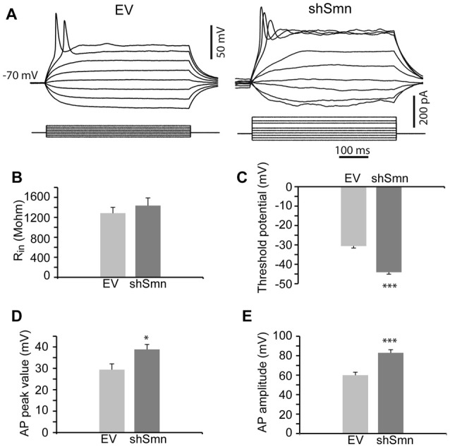
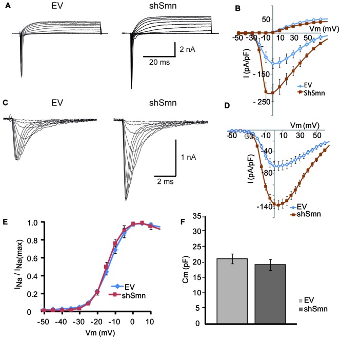
During development, motoneurons experience significant changes in their size and in the number and strength of connections that they receive, which requires adaptive changes in their passive and active electrical properties. Even after reaching maturity, motoneurons continue to adjust their intrinsic excitability and synaptic activity for proper functioning of the sensorimotor circuit in accordance with physiological demands. Likewise, if some elements of the circuit become dysfunctional, the system tries to compensate for the alterations to maintain appropriate function. In Spinal Muscular Atrophy (SMA), a severe motor disease, spinal motoneurons receive less excitation from glutamatergic sensory fibers and interneurons and are electrically hyperexcitable. Currently, the origin and relationship among these alterations are not completely established. In this study, we investigated whether Survival of Motor Neuron (SMN), the ubiquitous protein defective in SMA, regulates the excitability of motoneurons before and after the establishment of the synaptic contacts. To this end, we performed patch-clamp recordings in embryonic spinal motoneurons forming complex synaptic networks in primary cultures, and in differentiated NSC-34 motoneuron-like cells in the absence of synaptic contacts. Our results show that in both conditions, Smn-deficient cells displayed lower action potential threshold, greater action potential amplitudes, and larger density of voltage-dependent sodium currents than cells with normal Smn-levels. These results indicate that Smn participates in the regulation of the cell-autonomous excitability of motoneurons at an early stage of development. This finding may contribute to a better understanding of motoneuron excitability in SMA during the development of the disease.

Keywords: hyperexcitability; ion currents; motoneurons; spinal muscular atrophy (SMA); synapses.


Figures

Figure 1
Figure 1
Smn-deficient spinal motoneurons in culture establish less synapse contacts than empty vector (EV)-transduced neurons. (A) Embryonic mouse motoneurons in primary culture develop an extended neural network with numerous synaptic contacts at DIV12, both in EV and shSmn-transduced cultures, as visualized by confocal microscopic Z-projections. Axosomatic and axodendritic synapses appear as punta (red, anti-vGLUT2; green, anti-PSD95. Merged images are also shown. (B) Examples of immunofluorescence for Smn (red) and bassoon (white) at the soma of EV and shSmn-transduced motoneurons. (C) Western blot for Smn content in EV and shSmn-transduced cultures. (D) Comparison of the Smn fluorescence surface area, somatic total surface area, and number of bassoon spots in EV and shSmn-transduced cultures. The Smn signal is significantly decreased in Smn-deficient motoneurons (p = 0.008. two-tail unpair t-test). The mean number of bassoon spots is also low in Smn-deficient motoneurons (16.1 ± 1.6 spots; 48 neurons, three independent experiments) in comparison with motoneurons in EV cultures (23.4 ± 2.1 spots; 31 neurons, 31 neurons, three independent experiments; *p = 0.006, two-tail unpair t-test). Error bars represent SEM. Calibration bars: 20 μm.
Figure 2
Figure 2
Smn-deficient motoneurons are hyperexcitable. (A) Examples of passive and active responses under current clamp in control (EV) and shSmn-transduced motoneurons. Upon current injection, Smn-deficient motoneurons exhibited a higher discharge rate than EV motoneurons. (B) The input resistance of the Smn-deficient motoneurons shows an upward trend compared with that in the EV-transduced motoneurons. (C) Smn-deficient motoneurons show a reduced action potential threshold potential relative to EV-transduced cells (p = 0.005). (D,E) shSmn-transduced motoneurons have larger action potential amplitudes compared to controls (p = 0.04). Resting potential was set at −70 mV by small holding currents. Graph values are the mean of the action potential amplitude for Smn-reduced and control motoneurons of three independent experiments ± SEM (error bars). Asterisk indicates significant differences between data from the experiments using student’s t-test (*p < 0.05).
Figure 3
Figure 3
The sodium current density is increased in Smn-deficient motoneurons. (A) Examples of families of total whole-cell currents in EV and shSmn-transduced motoneurons. (B) I/V curves show an increase in inward current in Smn-deficient motoneurons (red symbols) compared to that in EV-transduced motoneurons (blue symbols). However, the size of the outward currents at the end of the pulse was not significantly different. Current amplitude was normalized to cell size by dividing the absolute value of the peak current by the membrane capacitance. (C,D) In the presence of potassium and calcium channel blockers, the peak sodium current was significantly larger in Smn-deficient cells than in EV-transduced cells. (E) Smn-deficient motoneurons have lower membrane electrical capacitance than control motoneurons (p = 0.03; n = 12).
Figure 4
Figure 4
EV- and shSmn transduced NSC-34 neurons. Confocal images of fixed and immunostained control (EV) and shSmn cultures at DIV3 showing cells with bipolar or pyramidal somata, relatively long neurites and growth cone-like structures (examples marked by arrows). Lentivirally transduced cells display direct green fluorescence (GFP). (A) Representative examples of Smn expression as assessed by immunostaining in EV and shSmn-transduced cells (central panel). (B,C) Immunoreactivity to vGlut (B, central panels), PSD95 (B, right panels), and vesicular acetylcholine transporter (vAChT) (C, right panel) was homogeneous and revealed the absence of synaptic contacts. Calibration bars: 20 μm.
Figure 5
Figure 5
Smn-deficient NSC-34 cells are hyperexcitable. (A) Examples of passive and active responses under current clamp in EV and shSmn-transduced cells. (B) The mean input resistance of the Smn-deficient cells was not significantly different from EV-transduced cells. (C) Smn-deficient cells show a reduced action potential threshold potential relative to EV-transduced cells (p < 0.0001; n = 24 and 29 cells). (D,E) shSmn-transduced cells have larger action potential amplitude compared to control motoneurons (p < 0.0001). Resting potential was set at −70 mV by small holding currents. Graph values are the mean of the action potential amplitude for Smn-reduced and control cells of three independent experiments ± SEM (error bars). Asterisk indicates significant differences between data from the experiments using student’s t-test (*p < 0.05; ***p < 0.0005).
Figure 6
Figure 6
The sodium current density is increased in Smn-deficient NSC-34 cells. (A) Examples of families of total whole-cell currents in EV and shSmn-transduced NSC-34 cells. (B) I/V curves show an increase in inward current in Smn-deficient neurons (red symbols) compared to that in EV-transduced neurons (blue symbols). However, the size of the outward currents at the end of the pulse was not significantly different. Current amplitude was normalized to cell size by dividing the absolute value of the peak current by the membrane capacitance. (C,D) In the presence of potassium and calcium channel blockers, the peak sodium current was significantly larger in Smn-deficient than in EV cells. (E) Voltage-dependence of the sodium current in control and Smn-deficient cells were not significantly different. (F) In Smn-deficient cells membrane electrical capacitance was similar to control (p = 0.03; n = 18).
Figure 7
Figure 7
Glutamate receptor blockers do not change the density of sodium and potassium currents in NSC-34 cells. (A–C) Examples of families of total whole-cell currents in NSC-34 cells (left) and I/V curves show no significant differences in outward or inward current amplitudes among cells recorded in the absence of drugs (A); n = 13, in the presence of APV (100 μM) + CNQX (20 μM) during the recording (B); n = 10, and incubated for 3 days with the glutamate blockers (C); n = 11. Current amplitudes were normalized to cell size by dividing the absolute value of the peak current by the membrane capacitance.

References

    1. Aptowicz C. O., Kunkler P. E., Kraig R. P. (2004). Homeostatic plasticity in hippocampal slice cultures involves changes in voltage-gated Na+ channel expression. Brain Res. 998, 155–163. 10.1016/j.brainres.2003.11.035 - DOI - PMC - PubMed
    1. Arnold W., McGovern V. L., Sanchez B., Li J., Corlett K. M., Kolb S. J., et al. (2016). The neuromuscular impact of symptomatic SMN restoration in a mouse model of spinal muscular atrophy. Neurobiol. Dis. 87, 116–123. 10.1016/j.nbd.2015.12.014 - DOI - PMC - PubMed
    1. Battle D. J., Lau C.-K., Wan L., Deng H., Lotti F., Dreyfuss G. (2006). The Gemin5 protein of the SMN complex identifies snRNAs. Mol. Cell 23, 273–279. 10.1016/j.molcel.2006.05.036 - DOI - PubMed
    1. Cashman N. R., Durham H. D., Blusztajn J. K., Oda K., Tabira T., Shaw I. T., et al. (1992). Neuroblastoma x spinal cord (NSC) hybrid cell lines resemble developing motor neurons. Dev. Dyn. 194, 209–221. 10.1002/aja.1001940306 - DOI - PubMed
    1. Crawford T. O., Pardo C. A. (1996). The neurobiology of childhood spinal muscular atrophy. Neurobiol. Dis. 3, 97–110. 10.1006/nbdi.1996.0010 - DOI - PubMed

LinkOut - more resources