

Molecular mechanisms of ampelopsin from Ampelopsis megalophylla induces apoptosis in HeLa cells
- PMID: 28928812
- PMCID: PMC5588129
- DOI: 10.3892/ol.2017.6520
Molecular mechanisms of ampelopsin from Ampelopsis megalophylla induces apoptosis in HeLa cells
Abstract
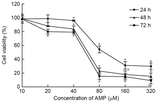
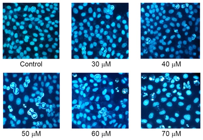
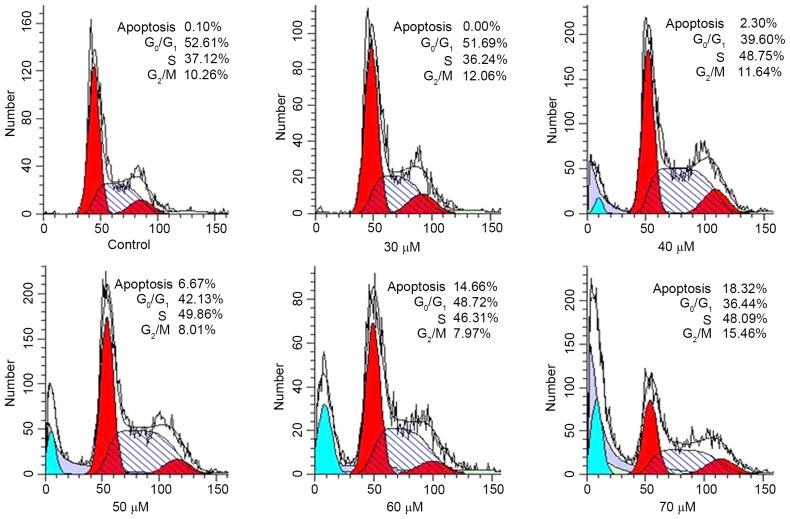
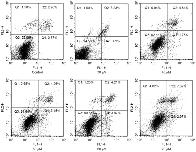
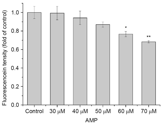
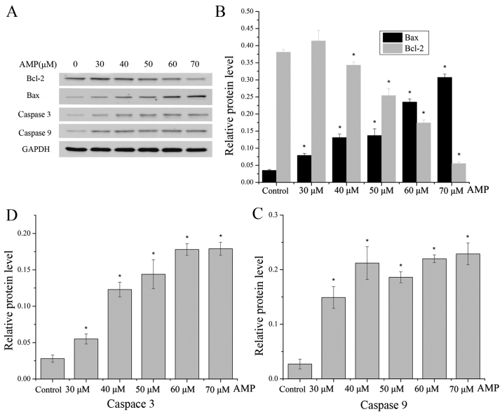
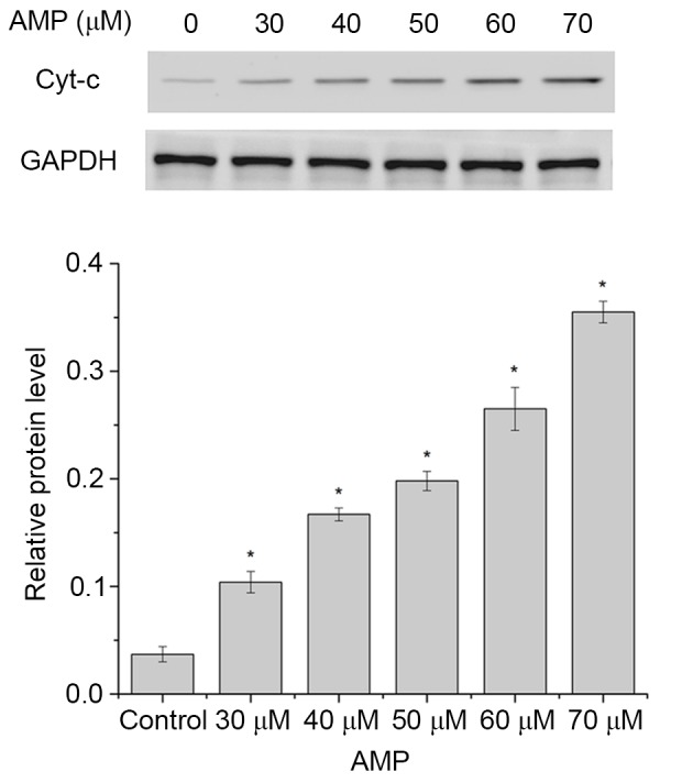
Ampelopsin (AMP) is an active ingredient of flavonoid compounds that is extracted from Ampelopsis megalophylla Diels et Gilg. The present study aimed at investigating the antitumor activities of AMP and the possible underlying molecular mechanisms in HeLa cells. A total of three types of tumor cell were selected to screen antitumor activities for AMP using the MTT assay. Flow cytometry was used to analyze the cell apoptotic proportion and the cell cycle. Rhodamine 123 staining was used to determine changes in mitochondrial transmembrane potential. Western blot analysis was used to determine the expression of apoptosis-associated proteins. The results of the present study demonstrated that AMP may inhibit the viability of HeLa cells in a dose- and time-dependent manner. Changes in morphology were observed using fluorescence microscopy. In addition, Annexin V-fluorescein isothiocyanate/propidium iodide (PI) double staining revealed that AMP induced apoptosis in a concentration-dependent manner and PI staining indicated that HeLa cells were arrested in S phase. Furthermore, western blot analysis demonstrated that AMP treatment induced apoptosis through activation of caspases 9 and 3, which was validated by the increasing ratio of B-cell lymphoma 2 (Bcl-2)-associated X protein to Bcl-2. Additionally, the loss of mitochondrial transmembrane potential and the release of cytochrome c suggested that AMP-induced apoptosis was associated with the mitochondrial pathway. Taken together, these results indicate that AMP may induce apoptosis via the mitochondrial signaling pathway in HeLa cells.
Keywords: Ampelopsis megalophylla Diels et Gilg; HeLa cells; ampelopsin; apoptosis; mitochondrial signaling pathway.
Figures
Similar articles
-
Total flavone extract from Ampelopsis megalophylla induces apoptosis in the MCF‑7 cell line.Int J Oncol. 2021 Mar;58(3):409-418. doi: 10.3892/ijo.2021.5172. Epub 2021 Jan 15. Int J Oncol. 2021. PMID: 33469684 Free PMC article.
-
Ampelopsin targets in cellular processes of cancer: Recent trends and advances.Toxicol Rep. 2022 Jul 27;9:1614-1623. doi: 10.1016/j.toxrep.2022.07.013. eCollection 2022. Toxicol Rep. 2022. PMID: 36561961 Free PMC article. Review.
-
Pseudolaric acid B exerts antitumor activity via suppression of the Akt signaling pathway in HeLa cervical cancer cells.Mol Med Rep. 2015 Aug;12(2):2021-6. doi: 10.3892/mmr.2015.3615. Epub 2015 Apr 15. Mol Med Rep. 2015. PMID: 25891953
-
Danthron, an anthraquinone derivative, induces DNA damage and caspase cascades-mediated apoptosis in SNU-1 human gastric cancer cells through mitochondrial permeability transition pores and Bax-triggered pathways.Chem Res Toxicol. 2011 Jan 14;24(1):20-9. doi: 10.1021/tx100248s. Epub 2010 Dec 2. Chem Res Toxicol. 2011. PMID: 21126053
-
Mitofusin-2 Triggers Cervical Carcinoma Cell Hela Apoptosis via Mitochondrial Pathway in Mouse Model.Cell Physiol Biochem. 2018;46(1):69-81. doi: 10.1159/000488410. Epub 2018 Mar 20. Cell Physiol Biochem. 2018. PMID: 29587277
Cited by
-
Total flavone extract from Ampelopsis megalophylla induces apoptosis in the MCF‑7 cell line.Int J Oncol. 2021 Mar;58(3):409-418. doi: 10.3892/ijo.2021.5172. Epub 2021 Jan 15. Int J Oncol. 2021. PMID: 33469684 Free PMC article.
-
Eriodictyol Inhibits Proliferation, Metastasis and Induces Apoptosis of Glioma Cells via PI3K/Akt/NF-κB Signaling Pathway.Front Pharmacol. 2020 Feb 25;11:114. doi: 10.3389/fphar.2020.00114. eCollection 2020. Front Pharmacol. 2020. PMID: 32158391 Free PMC article.
-
Multiple molecular and cellular mechanisms of the antitumour effect of dihydromyricetin (Review).Biomed Rep. 2024 Mar 26;20(5):82. doi: 10.3892/br.2024.1769. eCollection 2024 May. Biomed Rep. 2024. PMID: 38628627 Free PMC article. Review.
-
Ampelopsin targets in cellular processes of cancer: Recent trends and advances.Toxicol Rep. 2022 Jul 27;9:1614-1623. doi: 10.1016/j.toxrep.2022.07.013. eCollection 2022. Toxicol Rep. 2022. PMID: 36561961 Free PMC article. Review.
-
Dihydromyricetin Acts as a Potential Redox Balance Mediator in Cancer Chemoprevention.Mediators Inflamm. 2021 Mar 11;2021:6692579. doi: 10.1155/2021/6692579. eCollection 2021. Mediators Inflamm. 2021. PMID: 33776577 Free PMC article. Review.
References
LinkOut - more resources
Full Text Sources
Other Literature Sources
Research Materials
Miscellaneous