. 2017 Sep;14(3):2845-2851.
doi: 10.3892/ol.2017.6507. Epub 2017 Jun 30.

Experimental study of a novel tumstatin on C6 brain glioma in vitro

Affiliations

Experimental study of a novel tumstatin on C6 brain glioma in vitro

Pengguo Zhang et al. Oncol Lett. 2017 Sep.

Abstract

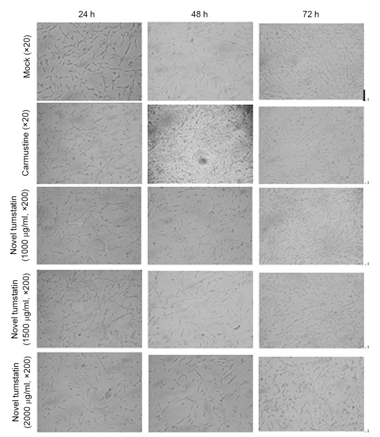
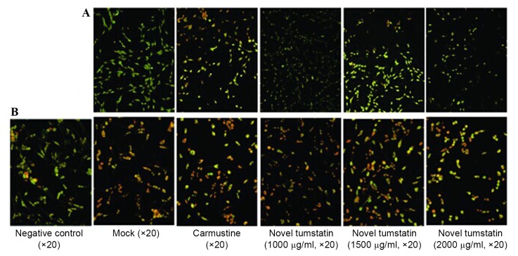
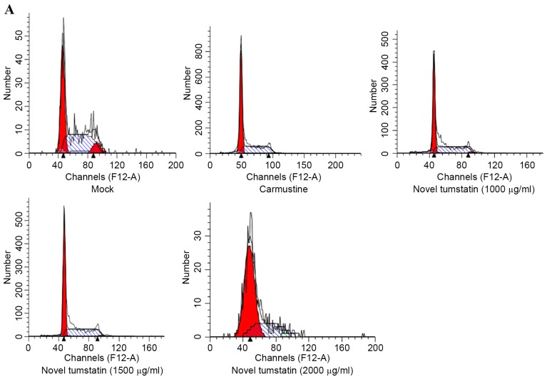
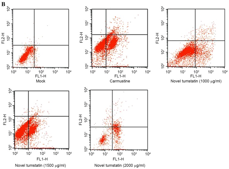
To investigate the effect of a novel tumstatin on C6 brain glioma cells, the MTT method was used to detect C6 glioma cell proliferation activity at different time periods (12, 48 and 72 h). Cell cycle distribution and apoptosis rate were detected by flow cytometry, and the acridine orange/ethidium bromide staining method was used to detect apoptosis and mitochondrial membrane potential by fluorescence microscopy. Novel tumstatin had an evident inhibitory effect on C6 glioma cells, and the most notable impact emerged after 48 h. The following were observed under the fluorescence microscope: Characteristic morphological changes of cell apoptosis were typically observed in the novel tumstatin (2,000 µg/ml) group; mitochondrial membrane potential decreased significantly (P<0.05); the cells in the G0/G1 phase significantly increased (P<0.05); and the number of cells in the S phase was reduced. There was an increase in cell apoptosis rate in the novel tumstatin (2,000 µg/ml) group compared with the novel tumstatin (1,000 µg/ml) group and the Mock group, and the data were statistically significant (P<0.05). Novel tumstatin may reduce the mitochondrial membrane potential, inducing cell apoptosis, and thereby exerting antitumor activity.

Keywords: C6 glioma cell; cell apoptosis; flow cytometry; novel tumstatin.


Figures

Figure 1.
Figure 1.
Inhibitory effects of different doses (0, 1,000, 1,500 and 2,000 µg/ml) of novel tumstatin on C6 glioma cells at 24, 48 and 72 h. The effects of novel tumstatin were determined by the MTT assay.
Figure 2.
Figure 2.
Apoptotic effects of novel tumstatin on the C6 cells after 48 h. (A) C6 glioma cells were treated by novel tumstatin for 48 h, using acridine orange/ethidium bromide staining for observation of cell apoptosis. (B) Mitochondrial membrane potential assay determined the apoptotic effect.
Figure 3.
Figure 3.
(A) Effects of different doses of novel tumstatin on the cell cycle of C6 glioma cells after 48 h. Cells were treated with different doses (0, 1,000, 1,500 and 2,000 µg/ml) of novel tumstatin and carmustine for 48 h, then analyzed by flow cytometry. (B) Effects of different doses of novel tumstatin on the cell apoptosis of C6 glioma cells after 48 h. Cells were treated with different doses (0, 1,000, 1,500 and 2,000 µg/ml) of novel tumstatin and carmustine for 48 h, and were then double labeled with Annexin V-fluorescein isothiocyanate/propidium iodide and analyzed by flow cytometry.
Figure 3.
Figure 3.
(A) Effects of different doses of novel tumstatin on the cell cycle of C6 glioma cells after 48 h. Cells were treated with different doses (0, 1,000, 1,500 and 2,000 µg/ml) of novel tumstatin and carmustine for 48 h, then analyzed by flow cytometry. (B) Effects of different doses of novel tumstatin on the cell apoptosis of C6 glioma cells after 48 h. Cells were treated with different doses (0, 1,000, 1,500 and 2,000 µg/ml) of novel tumstatin and carmustine for 48 h, and were then double labeled with Annexin V-fluorescein isothiocyanate/propidium iodide and analyzed by flow cytometry.

Similar articles

Cited by

References

    1. Auffinger B, Spencer D, Pytel P, Ahmed AU, Lesniak MS. The role of glioma stem cells in chemotherapy resistance and glioblastoma multiforme recurrence. Expert Rev Neurother. 2015;15:741–752. doi: 10.1586/14737175.2015.1051968. - DOI - PMC - PubMed
    1. Sancho-Martinez I, Martin-Villalba A. Tyrosine phosphorylation and CD95: A FAScinating switch. Cell Cycle. 2009;8:838–842. doi: 10.4161/cc.8.6.7906. - DOI - PubMed
    1. Eisele G, Roth P, Hasenbach K, Aulwurm S, Wolpert F, Tabatabai G, Wick W, Weller M. APO010, a synthetic hexameric CD95 ligand, induces human glioma cell death in vitro and in vivo. Neuro Oncol. 2011;13:155–164. doi: 10.1093/neuonc/noq176. - DOI - PMC - PubMed
    1. Konkankit VV, Kim W, Koya RC, Eskin A, Dam MA, Nelson S, Ribas A, Liau LM, Prins RM. Decitabine immunosensitizes human gliomas to NY-ESO-1 specific T lymphocyte targeting through the Fas/Fas ligand pathway. J Transl Med. 2011;9:192. doi: 10.1186/1479-5876-9-192. - DOI - PMC - PubMed
    1. Zhao JX, Liu XQ, Dong BJ. Primary study of bevacizumab combined with temozolomide for recurrent glioma. Chin Clin Oncol. 2011;39:920–922.