

Disrupted fibroblastic reticular cells and interleukin-7 expression in tumor draining lymph nodes
- PMID: 28928833
- PMCID: PMC5588138
- DOI: 10.3892/ol.2017.6537
Disrupted fibroblastic reticular cells and interleukin-7 expression in tumor draining lymph nodes
Abstract
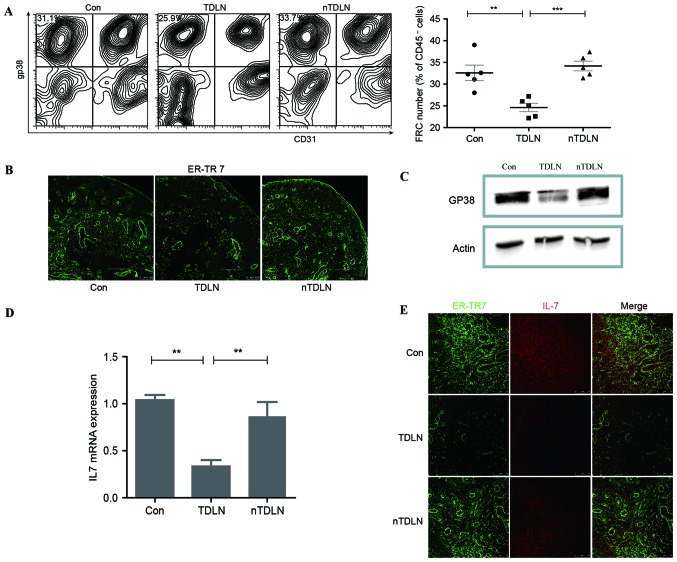
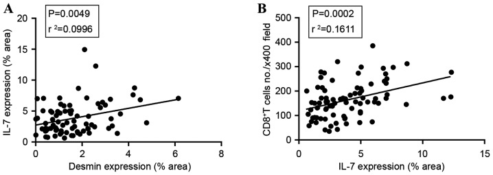
The immune system of patients with cancer is usually in an inhibitory state. Lymph node (LN) draining of pathological sites provides a suitable microenvironment where adaptive immune responses mainly occur. However, the microenvironment in the tumor draining lymph nodes (TDLNs) of patients with cancer appears to be in favor of tolerance. The effects of tumor cells on TDLNs have not been elaborated clearly. The present results have indicated that tumor cells may directly affect TDLNs by decreasing the fibroblastic reticular cell population that led to less interleukin-7 secretion. As a result, the number of T cells in TDLNs declined with reduced survival signals. A decreased number of T cells in TDLNs means weakened ability of immune surveillance. Clinically, these results were also confirmed in LN biopsies from patients with colon cancer at different clinical stages. Results of the present study showed that tumor cells may directly inhibit the immunological function of TDLNs.
Keywords: cancer; fibroblastic reticular cells; interleukin-7; tumor draining lymph node.
Figures


Similar articles
-
Single-cell analysis reveals immune modulation and metabolic switch in tumor-draining lymph nodes.Oncoimmunology. 2020 Oct 19;9(1):1830513. doi: 10.1080/2162402X.2020.1830513. Oncoimmunology. 2020. PMID: 33117603 Free PMC article.
-
Multi-scale characterization of tumor-draining lymph nodes in resectable lung cancer treated with neoadjuvant immune checkpoint inhibitors.EBioMedicine. 2022 Oct;84:104265. doi: 10.1016/j.ebiom.2022.104265. Epub 2022 Sep 15. EBioMedicine. 2022. PMID: 36116212 Free PMC article.
-
Unleashing the therapeutic potential of tumor-draining lymph nodes: spotlight on bladder cancer.J Transl Med. 2025 Apr 29;23(1):489. doi: 10.1186/s12967-024-05864-7. J Transl Med. 2025. PMID: 40301883 Free PMC article. Review.
-
Rectal cancer induces a regulatory lymphocytic phenotype in the tumor-draining lymph nodes to promote cancer cell installation.Immunol Res. 2020 Dec;68(6):363-372. doi: 10.1007/s12026-020-09161-5. Epub 2020 Nov 4. Immunol Res. 2020. PMID: 33150567
-
Tumor draining lymph nodes, immune response, and radiotherapy: Towards a revisal of therapeutic principles.Biochim Biophys Acta Rev Cancer. 2022 May;1877(3):188704. doi: 10.1016/j.bbcan.2022.188704. Epub 2022 Feb 25. Biochim Biophys Acta Rev Cancer. 2022. PMID: 35227831 Review.
Cited by
-
Tumor-draining lymph nodes: At the crossroads of metastasis and immunity.Sci Immunol. 2021 Sep 10;6(63):eabg3551. doi: 10.1126/sciimmunol.abg3551. Epub 2021 Sep 3. Sci Immunol. 2021. PMID: 34516744 Free PMC article. Review.
-
Progression of Metastasis through Lymphatic System.Cells. 2021 Mar 12;10(3):627. doi: 10.3390/cells10030627. Cells. 2021. PMID: 33808959 Free PMC article. Review.
-
Lymph Node Stromal Cells: Mapmakers of T Cell Immunity.Int J Mol Sci. 2020 Oct 21;21(20):7785. doi: 10.3390/ijms21207785. Int J Mol Sci. 2020. PMID: 33096748 Free PMC article. Review.
-
GFAP and desmin expression in lymphatic tissues leads to difficulties in distinguishing between glial and stromal cells.Sci Rep. 2021 Jun 25;11(1):13322. doi: 10.1038/s41598-021-92364-z. Sci Rep. 2021. PMID: 34172765 Free PMC article.
-
C-C Chemokine 21-Expressing T-cell Zone Fibroblastic Reticular Cells, Abundant in Lymph Nodes, Are Absent in Cancer Lymphoid Stroma.Acta Histochem Cytochem. 2024 Apr 25;57(2):67-74. doi: 10.1267/ahc.23-00066. Epub 2024 Apr 4. Acta Histochem Cytochem. 2024. PMID: 38695036 Free PMC article.
References
LinkOut - more resources
Full Text Sources
Other Literature Sources
Research Materials