

Twisted Gastrulation BMP Signaling Modulator 1 Regulates Papillary Thyroid Cancer Cell Motility and Proliferation
- PMID: 28928871
- PMCID: PMC5604214
- DOI: 10.7150/jca.18482
Twisted Gastrulation BMP Signaling Modulator 1 Regulates Papillary Thyroid Cancer Cell Motility and Proliferation
Abstract
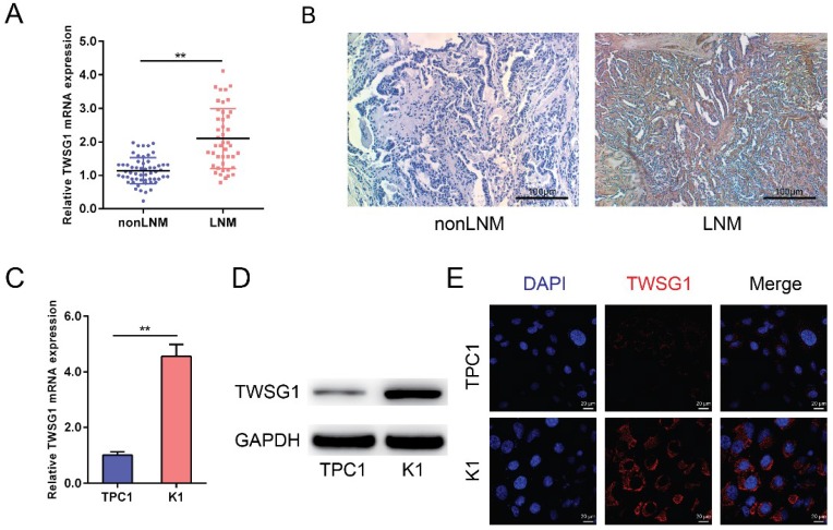
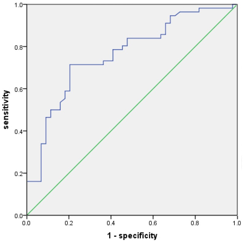
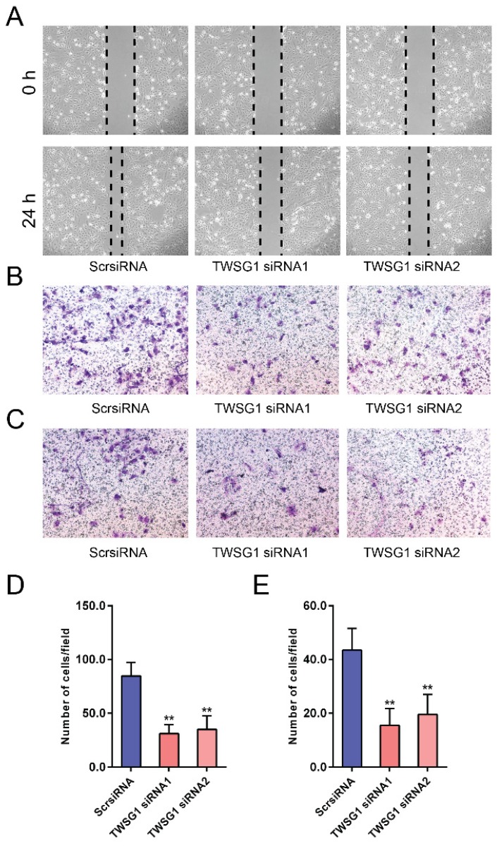
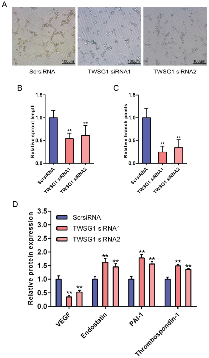
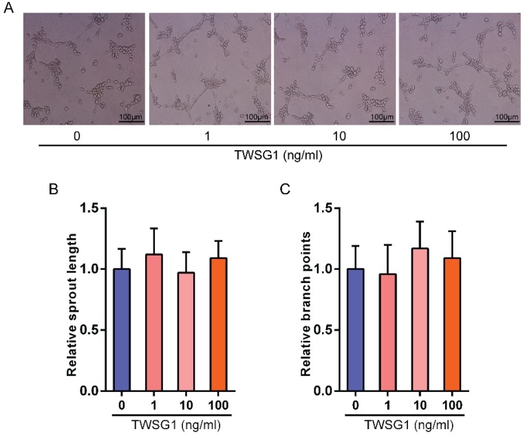
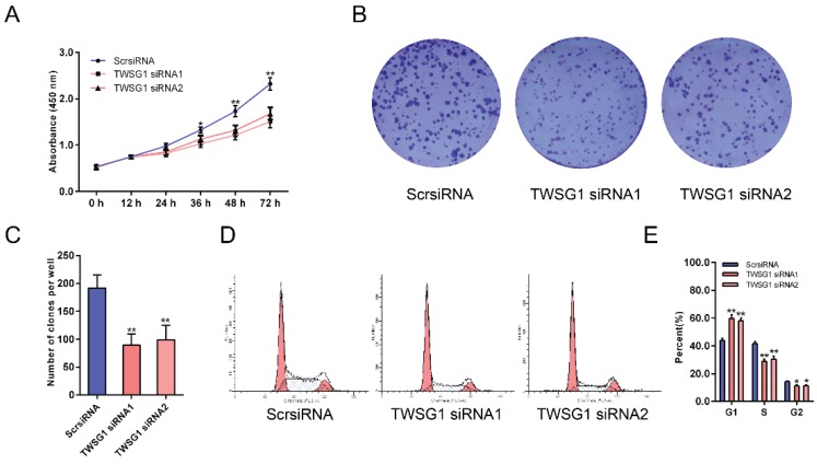
Bone morphogenetic proteins (BMPs) are growth factors that have important functions in cell proliferation, migration and differentiation. To date, BMP pathway activation has been found in multiple human tumors and is associated with enhanced malignant tumor growth and metastasis. BMP activity is tightly regulated by a family of soluble extracellular secreted BMP modulators. Twisted gastrulation BMP signaling modulator 1 (TWSG1) is a direct BMP regulator that is required for the full signaling activity of BMPs. However, the functions and mechanisms of TWSG1 in papillary thyroid cancer (PTC) metastasis have not been reported. TWSG1 expression was detected in 44 PTC tissues with lymph node metastasis (LNM) and 56 PTC tissues without LNM using quantitative real-time polymerase chain reaction (qRT-PCR). Gain- and loss-of-function approaches were used to assess the biological function of TWSG1 in PTC cells. Matrigel assays demonstrated the effect of tumor cell-derived TWSG1 on endothelial cell function. Our results showed that TWSG1 expression was significantly enhanced in PTC with LNM compared to that in PTC without LNM. TWSG1 knockdown inhibited migration, invasion and proliferation of PTC cells. Additionally, TWSG1 suppression impaired the tumor cell-induced endothelial cell sprout formation. We found that TWSG1 signaling may be transduced by the BMP target transcription factor inhibitor of DNA binding 1 (Id1) and matrix metalloproteinases (MMPs) 2 and 9. In conclusion, TWSG1 was highly expressed in metastasized PTC; tumor growth, migration and invasion were dependent on TWSG1, and it may be a new diagnostic and therapeutic target for PTC.
Keywords: TWSG1; biomarker.; invasion; migration; papillary thyroid cancer.
Conflict of interest statement
Competing Interests: The authors have declared that no competing interest exists.
Figures

Similar articles
-
Multifaceted Functions of TWSG1: From Embryogenesis to Cancer Development.Int J Mol Sci. 2022 Oct 22;23(21):12755. doi: 10.3390/ijms232112755. Int J Mol Sci. 2022. PMID: 36361543 Free PMC article. Review.
-
The Function of Twisted Gastrulation in Regulating Osteoclast Differentiation is Dependent on BMP Binding.J Cell Biochem. 2015 Oct;116(10):2239-46. doi: 10.1002/jcb.25174. J Cell Biochem. 2015. PMID: 25808976 Free PMC article.
-
Molecular mechanism of BMP signal control by Twisted gastrulation.Nat Commun. 2024 Jun 11;15(1):4976. doi: 10.1038/s41467-024-49065-8. Nat Commun. 2024. PMID: 38862520 Free PMC article.
-
BMP-binding protein twisted gastrulation is required in mammary gland epithelium for normal ductal elongation and myoepithelial compartmentalization.Dev Biol. 2013 Jan 1;373(1):95-106. doi: 10.1016/j.ydbio.2012.10.007. Epub 2012 Oct 24. Dev Biol. 2013. PMID: 23103586 Free PMC article.
-
Fibronectin 1 promotes migration and invasion of papillary thyroid cancer and predicts papillary thyroid cancer lymph node metastasis.Onco Targets Ther. 2017 Mar 22;10:1743-1755. doi: 10.2147/OTT.S122009. eCollection 2017. Onco Targets Ther. 2017. PMID: 28367057 Free PMC article.
Cited by
-
BMP signalling in colorectal cancer: losing the yin to WNTs yang.J Pathol. 2025 Jul;266(3):280-291. doi: 10.1002/path.6428. Epub 2025 Apr 11. J Pathol. 2025. PMID: 40212011 Free PMC article. Review.
-
The Role of Twisted Gastrulation 1 (TWSG1) Gene in TGF-β Signaling Linked to Cancer: A Comprehensive Review.Asian Pac J Cancer Prev. 2025 Apr 1;26(4):1129-1138. doi: 10.31557/APJCP.2025.26.4.1129. Asian Pac J Cancer Prev. 2025. PMID: 40302064 Free PMC article. Review.
-
Multifaceted Functions of TWSG1: From Embryogenesis to Cancer Development.Int J Mol Sci. 2022 Oct 22;23(21):12755. doi: 10.3390/ijms232112755. Int J Mol Sci. 2022. PMID: 36361543 Free PMC article. Review.
-
Hsa_circular RNA_0001013 exerts oncogenic effects in gastric cancer through the microRNA-136-TWSG1 axis.Am J Transl Res. 2022 Jul 15;14(7):4948-4963. eCollection 2022. Am J Transl Res. 2022. PMID: 35958507 Free PMC article.
-
Novel role of ASH1L histone methyltransferase in anaplastic thyroid carcinoma.J Biol Chem. 2020 Jun 26;295(26):8834-8845. doi: 10.1074/jbc.RA120.013530. Epub 2020 May 12. J Biol Chem. 2020. PMID: 32398261 Free PMC article.
References
-
- Davies L, Welch HG. Current thyroid cancer trends in the United States. JAMA otolaryngology- head & neck surgery. 2014;140:317–22. - PubMed
-
- Jemal A, Siegel R, Xu J, Ward E. Cancer statistics, 2010. CA: a cancer journal for clinicians. 2010;60:277–300. - PubMed
-
- Caron NR, Clark OH. Papillary thyroid cancer: surgical management of lymph node metastases. Current treatment options in oncology. 2005;6:311–22. - PubMed
LinkOut - more resources
Full Text Sources
Other Literature Sources
Miscellaneous
