Case Reports
. 2017:2017:9208474.
doi: 10.1155/2017/9208474. Epub 2017 Aug 8.

Long Standing Esophageal Perforation due to Foreign Body Impaction in Children: A Therapeutic Challenge in a Resource Limited Setting

Affiliations
Case Reports

Long Standing Esophageal Perforation due to Foreign Body Impaction in Children: A Therapeutic Challenge in a Resource Limited Setting

Ngo Nonga Bernadette et al. Case Rep Pediatr. 2017.

Abstract

Late presentation of foreign body impaction in the esophagus, complicated by perforation in children, has rarely been reported in the literature. Esophageal surgery is very difficult and challenging in Cameroon (a resource limited setting). We are reporting herein 2 cases of esophageal perforation in children seen very late (12 days and 40 days) after foreign body impaction, complicated with severe sepsis, who were successfully operated upon with very good results.


Figures

Figure 1
Figure 1
The type of impacted objects. A BIC pen and a 100 FCFA coin.
Figure 2
Figure 2
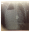
Chest X-ray of the boy at the time of the incident. The foreign body is visible in the chest. There is massive right pleural effusion and sign of esophageal perforation.
Figure 3
Figure 3
Chest X-ray one month later. This picture was taken the day of his arrival at our hospital. The empyema is loculated, the foreign body is still present in the chest, and the radiological signs of esophageal perforation are more obvious.
Figure 4
Figure 4
Surgical incision of the posterior thoracotomy in the baby boy one month after the surgery.
Figure 5
Figure 5
Chest X-ray, control one month after the surgery in the baby boy.
Figure 6
Figure 6
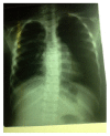
Chest X-ray of the 6 Y/O girl after the incident. Massive pleural effusion and air fluid level; foreign body in the right chest at the anatomical site of the esophagus and radiological signs of perforation.
Figure 7
Figure 7
Chest X-ray of the same girl 12 days after admission, loculated empyema, and foreign body at almost the same position.
Figure 8
Figure 8
(a) and (b) Esophagogram of the girl 2 months after surgery. There are no leaks. The esophagus appeared normal.
Figure 9
Figure 9
Chest X-ray: control at one year after both surgeries in the 6 Y/O girl.

References

    1. Ramareddy R. S., Alladi A. Review of esophageal injuries and stenosis: lessons learn and current concepts of management. Journal of Indian Association of Pediatric Surgeons. 2016;21(3):139–143. doi: 10.4103/0971-9261.182589. - DOI - PMC - PubMed
    1. Altokhais T. I., Al-Saleem A., Gado A., Al-Qahtani A., Al-Bassam A. Esophageal foreign bodies in children: Emphasis on complicated cases. Asian Journal of Surgery. 2015 doi: 10.1016/j.asjsur.2015.12.008. - DOI - PubMed
    1. Nonga B. N., Jemea B., Tambo F., Kamgaing N., Bahebeck J., Sosso M. A. Feasibility of a simple drainage system in Cameroonian children after thoracotomy and decortication for empyema thoracis. African Journal of Paediatric Surgery. 2012;9(1):27–31. doi: 10.4103/0189-6725.93298. - DOI - PubMed
    1. Alrazzak B., Elitsur Y., Al-Subu A. Etiology and management of esophageal impaction in children: a review of 11 years. Avicenna Journal of Medicine. 2013;3(2):33–36. doi: 10.4103/2231-0770.114113. - DOI - PMC - PubMed
    1. Tettey M., Edwin F., Aniteye E., et al. Management of intrathoracic oesophageal perforation: analysis of 16 cases. Tropical Doctor. 2011;41(4):201–203. doi: 10.1258/td.2011.110120. - DOI - PubMed

Publication types

LinkOut - more resources