. 2017 Oct 24;11(10):9738-9749.
doi: 10.1021/acsnano.7b01008. Epub 2017 Sep 20.

Heart Repair Using Nanogel-Encapsulated Human Cardiac Stem Cells in Mice and Pigs with Myocardial Infarction

Affiliations

Heart Repair Using Nanogel-Encapsulated Human Cardiac Stem Cells in Mice and Pigs with Myocardial Infarction

Junnan Tang et al. ACS Nano. .

Abstract

Stem cell transplantation is currently implemented clinically but is limited by low retention and engraftment of transplanted cells and the adverse effects of inflammation and immunoreaction when allogeneic or xenogeneic cells are used. Here, we demonstrate the safety and efficacy of encapsulating human cardiac stem cells (hCSCs) in thermosensitive poly(N-isopropylacrylamine-co-acrylic acid) or P(NIPAM-AA) nanogel in mouse and pig models of myocardial infarction (MI). Unlike xenogeneic hCSCs injected in saline, injection of nanogel-encapsulated hCSCs does not elicit systemic inflammation or local T cell infiltrations in immunocompetent mice. In mice and pigs with acute MI, injection of encapsulated hCSCs preserves cardiac function and reduces scar sizes, whereas injection of hCSCs in saline has an adverse effect on heart healing. In conclusion, thermosensitive nanogels can be used as a stem cell carrier: the porous and convoluted inner structure allows nutrient, oxygen, and secretion diffusion but can prevent the stem cells from being attacked by immune cells.

Keywords: biomaterials; cardiac stem cells; mouse model; myocardial infarction; nanogel; pig model.


Conflict of interest statement

The authors declare no competing financial interest.

Figures
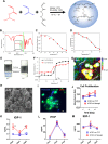
Figure 1
Figure 1
Synthesis of P(NIPAM-AA) nanogel and characterization of nanogel-encapsulated CSCs. (A) Schematic showing the synthesis of P(NIPAM-AA) nanogel by emulsion polymerization. (B) FTIR spectra of P(NIPAM-AA) thermoresponsive nanogel. (C) Temperature-dependent hydrodynamic diameter, dh, for 1 mg/mL of P(NIPAM-AA) nanogel in PBS. (D) Temperature-dependent shrinkage ratio dh(T)/dh (25 °C) in PBS. (E) Comparison of 30 mg/mL of P(NIPAM-AA) nanogel in PBS at sol state (25 °C) and gel state (37 °C). (F) Temperature-dependent dynamic rheological moduli of 30 mg/mL of P(NIPAM-AA) nanogel. Black closed circle corresponds to the elastic (or storage) modulus (G′), and the red circle corresponds to the viscous (or lose) modulus (G″). (G) Color-depth projection confocal image showing the morphology of CSCs encapsulated in the nanogel. Scale bar, 20 μm. (H) SEM image showing CSCs in the P(NIPAM-AA) nanogel. Scale bar, 20 μm. (I) Representative fluorescent image showing the morphology of CSCs cultured in nanogel. Scale bar, 10 μm. (J) Proliferation of CSCs cultured in P(NIPAM-AA) nanogel (red line) or on tissue culture plate (TCP) (blue line). (K–M) Release of insulin-like growth factor (IGF)-1, vascular endothelial growth factor (VEGF), and stromal cell-derived factor (SDF)-1 from hCSCs encapsulated in nanogel (red bar) or on TCP (blue bar) at various time points determined by ELISA; * indicates P < 0.05 when compared to the other group.
Figure 2
Figure 2
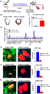
Impact of nanogel-encapsulated xenogeneic cardiac stem cells on cell retention and systemic inflammation in mice. (A) Schematic image indicating the general animal study design. (B) Ex vivo fluorescent imaging of mouse hearts at day 7 after injection of hCSCs in PBS or hCSCs in nanogel. (C) Quantitative PCR analysis of human cell retention in the mouse hearts (n = 3 animals per group). (D) Circulating levels of pro-inflammatory factors were remarkably elevated in mice treated with hCSCs in PBS compared to those treated with hCSCs encapsulated in polymer. (E–G) Fluorescent images revealing the presence of CD3+ T cells, CD8+ T cells, and CD68+ macrophage cells (green) in hearts injected with hCSCs (red) in PBS or nanogel at day 7 (n = 3 animals per group). Scale bar, 100 μm; * indicates P < 0.05.
Figure 3
Figure 3
Injection of nanogel-encapsulated human cardiac stem cells reduces myocardial apoptosis and promotes angiomyogenesis. (A) General design of animal study to explore the possible treatment of nanogel-encapsulated hCSCs in a mouse model of MI. (B) Fluorescent images of TUNEL+ apoptotic cells (red) in nanogel alone or hCSCs in nanogel-treated hearts at 3 weeks. (C) Quantitative analysis of TUNEL+ apoptotic cells (n = 3 animals per group). Scale bar, 100 μm. (D) Representative images revealing Ki67-positive cardiomyocyte nuclei (green) in nanogel or hCSCs in nanogel-treated hearts at 3 weeks. (E) Quantitative analysis of Ki67-positive nuclei both in scar zone and border zone (n = 3 hearts per group). Scale bar, 100 μm. (F) Representative images showing vWF-positive endothelial cells (green) in nanogel or hCSCs in nanogel-treated hearts at 3 weeks. Scale bar, 200 μm. (G) High-magnification image showing vessel formation (green) surrounding the injected nanogel-encapsulated CSCs (red). Scale bar, 50 μm. (H) Numbers of vWF-positive endothelial cells were quantified both in scar zone and border zone (n = 3 hearts per group); * indicates P < 0.05.
Figure 4
Figure 4
Injection of nanogel-encapsulated human cardiac stem cells augments cardiac function in a mouse model of MI. (A) Representative Masson’s trichrome-stained myocardial sections 3 weeks after treatment. (B–D) Quantitative analyses of viable myocardium (B), scar size (C), and infarct thickness (D) from the Masson’s trichrome images (n = 5 animals per group). (E,F) LVEFs determined by echocardiography at baseline (4 h post-MI) (E) and 3 weeks afterward (F) (n = 6 animals per group). (G) Treatment effects calculated as the change of LVEFs from baseline to end point; * indicates P < 0.05 when compared to “MI” group; # indicates P < 0.05 when compared to “MI + hCSCs” group; & indicates P < 0.05 when compared to “MI + nanogel” group.
Figure 5
Figure 5
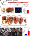
Nanogel encapsulation boosted cell retention in pig hearts. (A) Study design of the pig experiment. (B) Schematic images showing intramyocardial injection of nanogel-encapsulated CSCs in a pig heart. (C) Fluorescent micrograph and (D) quantitative analysis showing the presence of CD3+ T cells (red) in MI alone (white bar) or nanogel-encapsulated CSC (red bar)-treated hearts at 24 h (n = 3 animals per group). Scale bar, 100 μm. (E) Macroscopic images revealing infarct area on multiple slices of an infarcted pig heart. (F) Representative ex vivo fluorescent images and quantitative analysis of fluorescent intensities of pig hearts 24 h after injection of hCSCs in PBS (blue bar) or hCSCs in nanogel (red bar); * indicates P < 0.05.
Figure 6
Figure 6
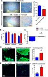
Nanogel-encapsulated CSC therapy reduces scar and preserves cardiac function in pigs with acute MI. (A) Featured Masson’s trichrome-stained myocardial sections 4 weeks after treatment in the infarct area and quantitative analysis of scar transmurality. (B) LVEFs determined by echocardiography at baseline (before infarct), post-MI (48 h post-infarct), and 4 weeks afterward. (C) Treatment effects calculated as the change of LVEFs from post-MI to end point. (D) Representative images indicating alpha-SA+ cardiomyocyte (green) in hearts treated with hCSCs in PBS or hCSCs in nanogel (n = 3 hearts per group) at 4 weeks. Quantitative analysis of alpha-SA+ cardiomyocyte. Scale bar, 100 μm. (E) Representative images exhibiting alpha-SMA+ vasculatures (green) in hearts treated with hCSCs in PBS or hCSCs in nanogel (n = 3 hearts per group) at 4 weeks. The numbers of alpha-SMA+ vasculatures were quantified. Scale bar, 200 μm; * indicates P < 0.05.

References

    1. Bolli R.; Chugh A. R.; D’Amario D.; Loughran J. H.; Stoddard M. F.; Ikram S.; Beache G. M.; Wagner S. G.; Leri A.; Hosoda T.; et al. Cardiac Stem Cells in Patients with Ischaemic Cardiomyopathy (SCIPIO): Initial Results of a Randomised Phase 1 Trial. Lancet 2011, 378, 1847–57. 10.1016/S0140-6736(11)61590-0. - DOI - PMC - PubMed
    1. Sayed N.; Liu C.; Wu J. C. Translation of Human-Induced Pluripotent Stem Cells: From Clinical Trial in a Dish to Precision Medicine. J. Am. Coll. Cardiol. 2016, 67, 2161–76. 10.1016/j.jacc.2016.01.083. - DOI - PMC - PubMed
    1. Hunsberger J. G.; Rao M.; Kurtzberg J.; Bulte J. W.; Atala A.; LaFerla F. M.; Greely H. T.; Sawa A.; Gandy S.; Schneider L. S.; et al. Accelerating Stem Cell Trials for Alzheimer’s Disease. Lancet Neurol. 2016, 15, 219–230. 10.1016/S1474-4422(15)00332-4. - DOI - PubMed
    1. Bartunek J.; Davison B.; Sherman W.; Povsic T.; Henry T. D.; Gersh B.; Metra M.; Filippatos G.; Hajjar R.; Behfar A.; et al. Congestive Heart Failure Cardiopoietic Regenerative Therapy (CHART-1) Trial Design. Eur. J. Heart Failure 2016, 18, 160–8. 10.1002/ejhf.434. - DOI - PMC - PubMed
    1. van Berlo J. H.; Molkentin J. D. An Emerging Consensus on Cardiac Regeneration. Nat. Med. 2014, 20, 1386–93. 10.1038/nm.3764. - DOI - PMC - PubMed

Publication types