. 2017 Sep 13;18(Suppl 10):402.
doi: 10.1186/s12859-017-1788-4.

Reconstruction and visualization of large-scale volumetric models of neocortical circuits for physically-plausible in silico optical studies

Affiliations

Reconstruction and visualization of large-scale volumetric models of neocortical circuits for physically-plausible in silico optical studies

Marwan Abdellah et al. BMC Bioinformatics. .

Abstract

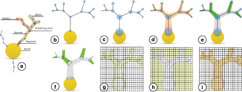
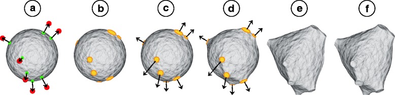
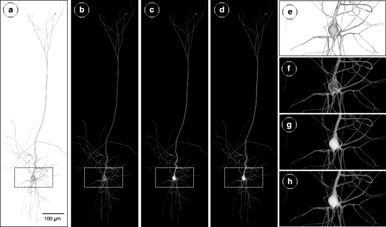
Background: We present a software workflow capable of building large scale, highly detailed and realistic volumetric models of neocortical circuits from the morphological skeletons of their digitally reconstructed neurons. The limitations of the existing approaches for creating those models are explained, and then, a multi-stage pipeline is discussed to overcome those limitations. Starting from the neuronal morphologies, we create smooth piecewise watertight polygonal models that can be efficiently utilized to synthesize continuous and plausible volumetric models of the neurons with solid voxelization. The somata of the neurons are reconstructed on a physically-plausible basis relying on the physics engine in Blender.

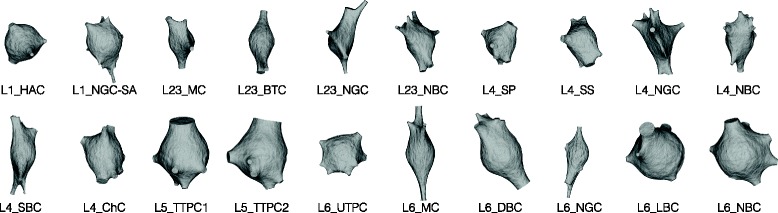
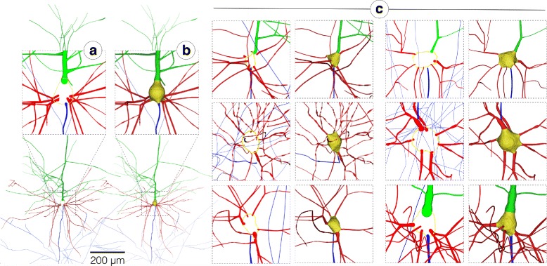
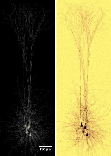
Results: Our pipeline is applied to create 55 exemplar neurons representing the various morphological types that are reconstructed from the somatsensory cortex of a juvenile rat. The pipeline is then used to reconstruct a volumetric slice of a cortical circuit model that contains ∼210,000 neurons. The applicability of our pipeline to create highly realistic volumetric models of neocortical circuits is demonstrated with an in silico imaging experiment that simulates tissue visualization with brightfield microscopy. The results were evaluated with a group of domain experts to address their demands and also to extend the workflow based on their feedback.

Conclusion: A systematic workflow is presented to create large scale synthetic tissue models of the neocortical circuitry. This workflow is fundamental to enlarge the scale of in silico neuroscientific optical experiments from several tens of cubic micrometers to a few cubic millimeters.

Ams subject classification: Modelling and Simulation.

Keywords: In silico neuroscience; Modeling and simulation; Neocortical brain models; Polygonal and volumetric models.


Conflict of interest statement

Ethics approval and consent to participate

Not applicable.

Consent for publication

Not applicable.

Competing interests

The authors declare that they have no competing interests.

Publisher’s Note

Springer Nature remains neutral with regard to jurisdictional claims in published maps and institutional affiliations.

Figures

Fig. 1
Fig. 1
An illustration of our proposed workflow for creating volumetric models of the neurons from their morphological skeletons. a A graphical representation of a typical morphological skeleton of a neuron. To eliminate any visual distractions, the workflow will be illustrated using a single arbor sampled only at the branching points (b-f). The blue circles in b and c represent the positions of morphological samples of the neurons and the radii of their respective cross-sections. d The morphology structure is created by connecting the samples, segments, and branches together. e The primary branches that represent a continuation along the arbor (in the same color) are identified according to the radii of samples of the children branches at the bifurcation points. f The connected branches identified in (e) are converted into multiple mesh objects where each object is smooth and watertight. g The mesh objects are converted to intersecting volumetric shells with surface voxelization in the same volume. h Solid voxelization. The volume created in (g) is flood-filled to cover the extra-cellular space of the neurons. i The final volumetric model of a neuron is created by inverting the flood-filled volume to reflect a smooth, continuous and plausible representation of the neuron
Fig. 2
Fig. 2
Soma progressive reconstruction. The soma is modeled by a soft body sphere in (a). The initial and final locations of the primary branches are illustrated by the green and red points respectively. The first-order sections are projected to the sphere to find out the vertices where the hooks will be created. The faces from each hook are merged into a single face and shaped into a circle (b). The hooks are pulled and the circles are scaled to match the size of the sections (c-e). The final soma is reconstructed in (f)
Fig. 3
Fig. 3
Physically-plausible reconstruction of the somata of diverse neocortical neurons labeled by their morphological type. The initial shape of the soma is defined by a soft body sphere that is deformed by pulling the corresponding vertices of each primary branch. The algorithm uses the soft body toolbox and the hook modifier in Blender [30]
Fig. 4
Fig. 4
Reconstruction of a piecewise watertight polygonal mesh model of a pyramidal neuron in (b) from its morphological skeleton in (a). In (c), the applicability of the proposed meshing algorithm is demonstrated with multiple neurons having diverse morphological types to validate its generality. The reconstruction results of the 55 exemplar neurons are provided in high resolution with the Additional file 1. The somata, basal dendrites, apical dendrites and axons are colored in yellow, red, green and blue respectively
Fig. 5
Fig. 5
The process of building a volumetric model of a single pyramidal neuron from its polygonal mesh. The polygonal mesh model in (a) is converted to a volumetric shell with surface voxelization in (b) and a filled volume with solid voxelization in (c). In (d), the spines are integrated to the volume. The images in (e), (f), (g) and (h) are close ups for the renderings in (a), (b), (c) and (d) respectively. Notice the overlapping shells of the different branches and the soma that result due to the surface voxelization step in (f). In (g), the volume created with solid voxelization reflects a continuous, smooth and high fidelity representation of the entire neuron
Fig. 6
Fig. 6
Volumetric reconstructions of multiple neocortical circuits with solid voxelization. The presented workflow is capable of creating large scale volumetric models for circuits with different complexity. a Single cell volume. b A group of five pyramidal neurons. c 5% of the pyramidal neurons that exist in layer five in the neocortical column. d 5% of all the neurons in a single column (containing ∼31,000 neurons). e A uniformly-sampled selection of only 1% of the neurons in a digital slice composed of seven columns (containing ∼210,000 neurons) stacked together. The resolution of the largest dimension of each volume is set to 8000 voxels. The area covered by the orange box in (e) represents the maximum volumetric extent that could be simulated in similar previous studies [28, 29]
Fig. 7
Fig. 7
In silico imaging of neuronal tissue with brightfield microscope. The volumetric model (left) is annotated with the optical characteristics of Golgi’s silver stain. The in silico image created in (b) is used to study the visual response of different dyes used in the in vitro experiment

References

    1. Koch C, Segev I. Methods in Neuronal Modeling: from Ions to Networks. Cambridge: MIT press; 1998.
    1. Geschwind DH, Konopka G. Neuroscience in the era of functional genomics and systems biology. Nature. 2009;461(7266):908–15. doi: 10.1038/nature08537. - DOI - PMC - PubMed
    1. Di Ventura B, Lemerle C, Michalodimitrakis K, Serrano L. From in vivo to in silico biology and back. Nature. 2006;443(7111):527–33. doi: 10.1038/nature05127. - DOI - PubMed
    1. Markram H. The Blue Brain Project. Nat Rev Neurosci. 2006;7(2):153–60. doi: 10.1038/nrn1848. - DOI - PubMed
    1. Sanbonmatsu K, Tung CS. High performance computing in biology: multimillion atom simulations of nanoscale systems. J Struct Biol. 2007;157(3):470–80. doi: 10.1016/j.jsb.2006.10.023. - DOI - PMC - PubMed

LinkOut - more resources