Fishing for Answers in Human Mycobacterial Infections
- PMID: 28930653
- PMCID: PMC6481165
- DOI: 10.1016/j.immuni.2017.09.005
Fishing for Answers in Human Mycobacterial Infections
Abstract
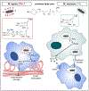
Two recent studies (Cambier et al., 2017; Madigan et al., 2017) reveal in vivo functions for specific phenolic glycolipids (PGLs) in the mycobacteria that cause tuberculosis or leprosy. M. tuberculosis (and M. marinum) PGL promotes bacterial spread to growth-permissive macrophages, while M. leprae PGL-1 induces macrophages to cause nerve demyelination characteristic of human leprosy.
Copyright © 2017 Elsevier Inc. All rights reserved.
Figures

Comment on
-
A Macrophage Response to Mycobacterium leprae Phenolic Glycolipid Initiates Nerve Damage in Leprosy.Cell. 2017 Aug 24;170(5):973-985.e10. doi: 10.1016/j.cell.2017.07.030. Cell. 2017. PMID: 28841420 Free PMC article.
-
Phenolic Glycolipid Facilitates Mycobacterial Escape from Microbicidal Tissue-Resident Macrophages.Immunity. 2017 Sep 19;47(3):552-565.e4. doi: 10.1016/j.immuni.2017.08.003. Epub 2017 Aug 24. Immunity. 2017. PMID: 28844797 Free PMC article.
References
-
- Hirsch CS, Ellner JJ, Russell DG, and Rich EA (1994). J. Immunol 152, 743–753. - PubMed
Publication types
MeSH terms
Substances
Grants and funding
LinkOut - more resources
Full Text Sources
Other Literature Sources
Miscellaneous

