

Accessory spleen located in the right parietal peritoneum: The first case report
- PMID: 28930831
- PMCID: PMC5617698
- DOI: 10.1097/MD.0000000000007957
Accessory spleen located in the right parietal peritoneum: The first case report
Abstract
Rationale: Accessory spleen is a congenital abnormality caused by failure of the splenic anlage to fuse during embryology. The presence of an accessory spleen located in the parietal peritoneum has not been reported so far, and an accessory spleen situated on the right side is extremely rare. In the present study, we describe the first case of an accessory spleen located in the right parietal peritoneum.
Patients concerns: A 65-year-old man, presented with pain in his left abdomen for 1 month.
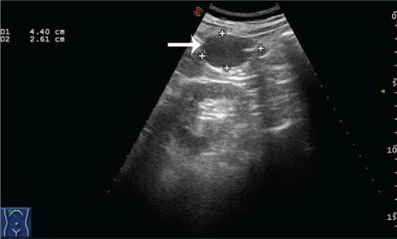
Diagnoses: With ultrasonography and computed tomography, it was difficult to determine the accurate location and diagnosis, and an abdominal fibroma was preoperatively considered.
Interventions: By laparotomy, we found a mass connected to the right parietal peritoneum by a vascular pedicle. We resected it completely, and the gross specimen measured 5.0 × 3.0 × 2.5 cm and was a localized tumor with a capsule.
Outcomes: Microscopically, sinusoids were visible, as well as scattered lymphoid follicles, eosinophils, histiocytes, plasma cells, neutrophils, and red blood cells, indicative of splenic tissue. Finally, the lesion was diagnosed as an accessory spleen located in the right parietal peritoneum. Postoperatively, he recovered well and was followed up for a 31 months, during which he was well with no complication.
Lessons: We present the first accessory spleen located in the right parietal peritoneum. Awareness of the accessory spleen and familiarity with typical imaging findings are necessary for surgeons to make a precise preoperative diagnosis.
Conflict of interest statement
The authors report no conflicts of interest.
Figures


Similar articles
-
Accessory spleen in the greater omentum.Am J Surg. 2011 Sep;202(3):e28-30. doi: 10.1016/j.amjsurg.2010.06.032. Epub 2011 Jul 23. Am J Surg. 2011. PMID: 21784408
-
Evolution of the CT imaging findings of accessory spleen infarction.Pediatr Radiol. 2006 Dec;36(12):1319-22. doi: 10.1007/s00247-006-0323-y. Epub 2006 Oct 3. Pediatr Radiol. 2006. PMID: 17016699
-
The real cause of right lower abdominal pain: an analysis of ultrasonographic findings.J Int Med Res. 2020 Aug;48(8):300060520946185. doi: 10.1177/0300060520946185. J Int Med Res. 2020. PMID: 32841582 Free PMC article.
-
Colonic obstruction caused by accessory spleen torsion: A rare case report and literature review.Medicine (Baltimore). 2017 Sep;96(39):e8116. doi: 10.1097/MD.0000000000008116. Medicine (Baltimore). 2017. PMID: 28953636 Free PMC article. Review.
-
Accessory Spleen Fracture: Report of a Pediatric Case and Review of the Literature.Pediatr Emerg Care. 2020 Jan;36(1):e10-e13. doi: 10.1097/PEC.0000000000001381. Pediatr Emerg Care. 2020. PMID: 29298250 Review.
Cited by
-
Expanding Role of Contrast-Enhanced Ultrasound and Elastography in the Evaluation of Abdominal Pathologies in Children.Diagnostics (Basel). 2025 Jul 1;15(13):1680. doi: 10.3390/diagnostics15131680. Diagnostics (Basel). 2025. PMID: 40647679 Free PMC article. Review.
-
An extremely rare case of an oversized accessory spleen: case report and review of the literature.BMC Surg. 2019 Apr 27;19(1):45. doi: 10.1186/s12893-019-0510-z. BMC Surg. 2019. PMID: 31029135 Free PMC article. Review.
-
Infarcted accessory spleen masquerading as a mesenteric cyst.BMJ Case Rep. 2018 Aug 16;2018:bcr2018226130. doi: 10.1136/bcr-2018-226130. BMJ Case Rep. 2018. PMID: 30115724 Free PMC article.
References
-
- Wadham BM, Adams PB, Johnson MA. Incidence and location of accessory spleens. N Engl J Med 1981;304:1111. - PubMed
-
- Halpert B, Gyorkey F. Lesions observed in accessory spleens of 311 patients. Am J Clin Pathol 1959;32:165–8. - PubMed
-
- Leon L, Labropoulos N, Hudlin CI, et al. Accessory spleen rupture in a patient with previous traumatic splenectomy. J Trauma 2006;60:901–3. - PubMed
-
- Halpert B, Alden ZA. Accessory spleens in or at the tail of the Pancreas. A survey of 2,700 additional necropsies. Arch Pathol 1964;77:652–4. - PubMed
Publication types
MeSH terms
LinkOut - more resources
Full Text Sources
Other Literature Sources
Medical
