Comparative Study
. 2017 Sep 20;12(9):e0185028.
doi: 10.1371/journal.pone.0185028. eCollection 2017.

miR-181a decelerates proliferation in cutaneous squamous cell carcinoma by targeting the proto-oncogene KRAS

Affiliations
Comparative Study

miR-181a decelerates proliferation in cutaneous squamous cell carcinoma by targeting the proto-oncogene KRAS

Johannes Neu et al. PLoS One. .

Abstract

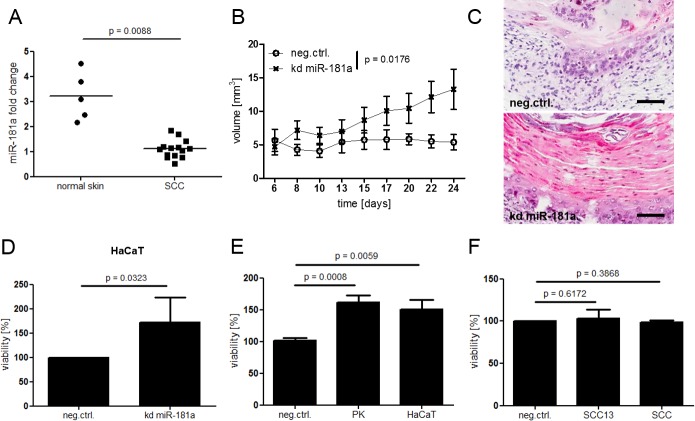
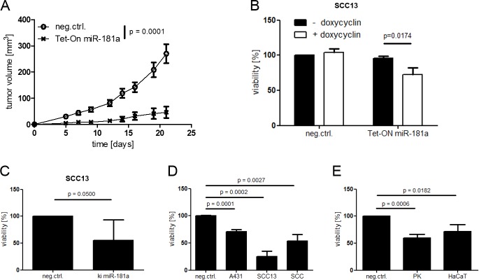
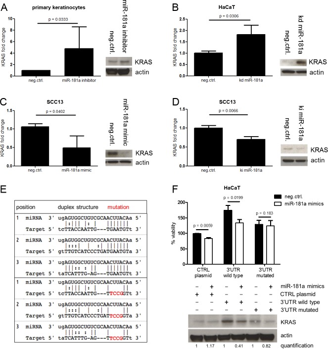
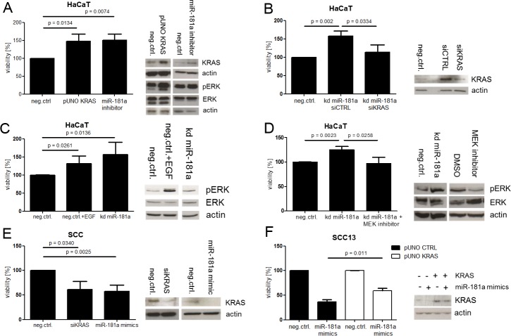
Cutaneous squamous cell carcinoma (SCC) is the second most common human skin cancer with a rapidly increasing incidence among the Caucasian population. Among the many regulators, responsible for cancer progression and growth, microRNAs (miRNA) are generally accepted as key players by now. In our current study we found that microRNA-181a (miR-181a) shows low abundance in SCC compared to normal epidermal skin. In vitro, miRNA downregulation in normal primary keratinocytes induced increased proliferation, while in vivo miR-181a downregulation in HaCaT normal keratinocytes showed tumor-like growth increase up to 50%. Inversely, upregulation of these miRNAs in cancer cells lead to reduced cellular proliferation and induction of apoptosis in vitro. An in vivo therapeutic model with induced miR-181a expression in SCC13 cancer cells reduced tumor formation in mice by 80%. Modulation of miR-181a levels showed an inverse correlation with the proto-oncogene KRAS both on mRNA and protein level by direct interaction. Knockdown of KRAS mimicked the anti-proliferative effects of miR-181a overexpression in patient-derived SCC cells and abolished the enhanced viability of HaCaT cells following miR-181a knockdown. Furthermore, phospho-ERK levels correlated with KRAS levels, suggesting that the observed effects were mediated via the MAPK signaling pathway. miR-181a seemed regulated during keratinocyte differentiation probably in order to amplify the tumor suppressive character of differentiation. Taken together, miR-181a plays a crucial tumor suppressive role in SCC by targeting KRAS and could be a promising candidate for a miRNA based therapy.


Conflict of interest statement

Competing Interests: The authors have declared that no competing interests exist.

Figures

Fig 1
Fig 1. Low abundance of miR-181a is found in patient derived SCC specimen compared to normal skin and down regulation of miR-181a leads to increased viability in healthy keratinocytes.
(A) Total RNA was isolated from patient derived SCC or normal skin biopsies (epidermal part). miR-181a levels were determined via TaqMan qPCR. (B) HaCaT knock down (kd) miR-181a or control cells were injected subcutaneously into nude mice. Over all significance of the time course experiment was calculated using two way Anova and Bonferroni correction. A Student’s t-Test was performed at the end point (day 24) (Figure B in S4 Fig). (C) H&E sections of cysts. Black bar = 100μm. (D) Viability of HaCaT kd miR-181a cells in vitro. (E + F) Cells were transfected with miR-181a inhibitors for 48 hours. For in vitro viability assays cells were seeded into 96 well plates and incubated for 96 hours followed by WST-1 assay. P values for in vitro assays were Calculated via Student’s T test. SCC = cultured SCC cells derived from patient samples, PK = cultured healthy keratinocytes derived from patient samples.
Fig 2
Fig 2. Up regulation of miR-181a leads to decreased viability in cancer cells.
(A) SCC13 was transduced with pTRIPZ for inducible miRNA overexpression (SCC13 Tet-ON miR-181a) and injected subcutaneously into nude mice. Doxycycline (200 mg/kg) was administered via food pellets. Over all significance of the time course experiment was calculated using two way Anova and Bonferroni correction. (B) SCC13 Tet-On miR-181a were seeded into 96 well plates and exposed to doxycycline during the course of the experiment. Cells were transduced with pLKO.miRNA (ki miR-181a) for stable miRNA overexpression (C) or transfected with miRNA mimics for 48 hours (D + E). Cells were seeded into 96 well plates and incubated for 96 hours followed by WST-1 viability assay. P values for in vitro assays were calculated via Student’s t-Test. SCC = cultured SCC cells derived from patient samples, PK = cultured healthy keratinocytes derived from patient samples.
Fig 3
Fig 3. miR-181a targets KRAS directly.
Cells were transfected with miRNA mimics (A) or inhibitors (C) for 48 hours or transduced with pLKO.miRNA.inhibitor / pLKO.miRNA (kd miR-181a / ki miR-181a) for stable miRNA modulation (B and D). Cells were lysed and mRNA or protein level analyses were performed using SYBR green qPCR or Western Blot respectively. (F) HaCaT cells were stably transfected with the indicated pUNO KRAS over expression plasmids in selection medium. Selected clones were transfected with miR-181a mimics for 48 hours following by WST-1 assay and Western Blot. (E) The panel in the left illustrates the three main miR-181a binding sites in KRAS 3’UTR and the according mutations. Student’s t-Test was used to calculate P values.
Fig 4
Fig 4. miR-181a mediates its tumor suppressive role through KRAS which signals via the MAPK pathway.
Cells were transfected with the indicated siRNAs or miRNA mimics (A). In addition to siRNA/mimics the cells were transduced with the indicated plasmids (A—F). All transfections were carried out for 48 hours. A subset of the cells was seeded into 96 well plates, followed by WST-1 assay, while RNA and protein was isolated from the left overs. Protein and mRNA levels were determined by Western Blot or SYBR Green qPCR respectively. Student’s t-Test was used to calculate P values. kd = knock down, SCC = cultured SCC cells derived from patient samples.

Similar articles

Cited by

References

    1. Hofbauer GF, Bouwes Bavinck JN, Euvrard S. Organ transplantation and skin cancer: basic problems and new perspectives. Experimental dermatology. 2010;19(6):473–82. Epub 2010/05/21. doi: 10.1111/j.1600-0625.2010.01086.x . - DOI - PubMed
    1. Lohmann CM, Solomon AR. Clinicopathologic variants of cutaneous squamous cell carcinoma. Advances in anatomic pathology. 2001;8(1):27–36. Epub 2001/01/11. . - PubMed
    1. Ratushny V, Gober MD, Hick R, Ridky TW, Seykora JT. From keratinocyte to cancer: the pathogenesis and modeling of cutaneous squamous cell carcinoma. The Journal of clinical investigation. 2012;122(2):464–72. Epub 2012/02/02. doi: 10.1172/JCI57415 ; PubMed Central PMCID: PMCPMC3266779. - DOI - PMC - PubMed
    1. Brash DE, Rudolph JA, Simon JA, Lin A, McKenna GJ, Baden HP, et al. A role for sunlight in skin cancer: UV-induced p53 mutations in squamous cell carcinoma. Proceedings of the National Academy of Sciences of the United States of America. 1991;88(22):10124–8. Epub 1991/11/15. ; PubMed Central PMCID: PMCPMC52880. - PMC - PubMed
    1. Euvrard S, Kanitakis J, Claudy A. Skin cancers after organ transplantation. The New England journal of medicine. 2003;348(17):1681–91. Epub 2003/04/25. doi: 10.1056/NEJMra022137 . - DOI - PubMed

Publication types

MeSH terms