

Heterogeneity in the expression and subcellular localization of POLYOL/MONOSACCHARIDE TRANSPORTER genes in Lotus japonicus
- PMID: 28931056
- PMCID: PMC5607196
- DOI: 10.1371/journal.pone.0185269
Heterogeneity in the expression and subcellular localization of POLYOL/MONOSACCHARIDE TRANSPORTER genes in Lotus japonicus
Abstract
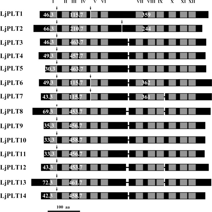
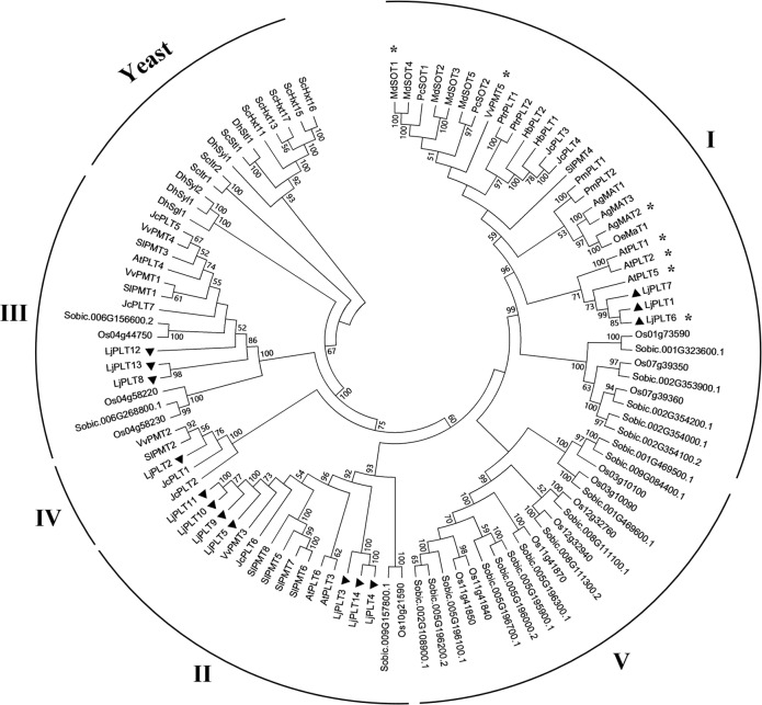
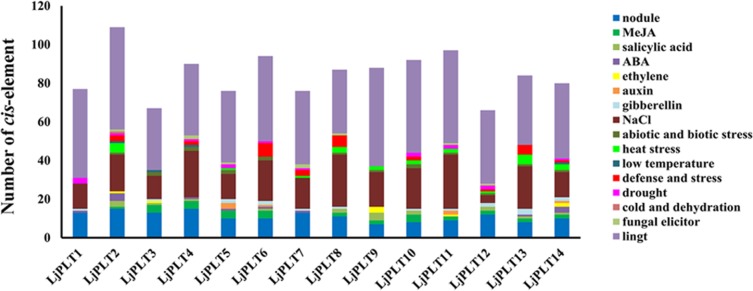
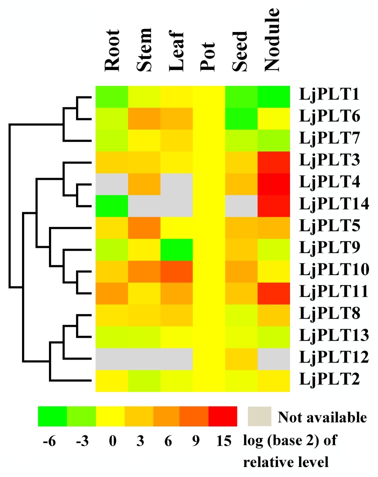
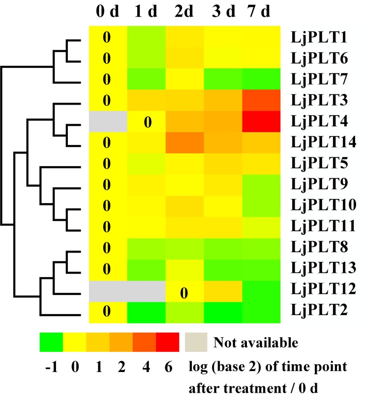
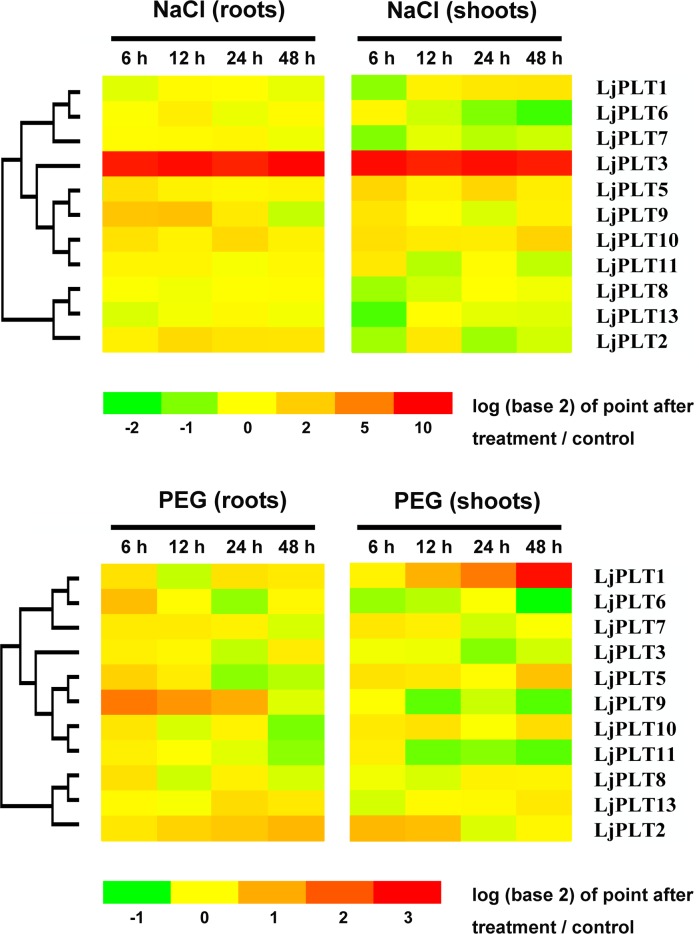
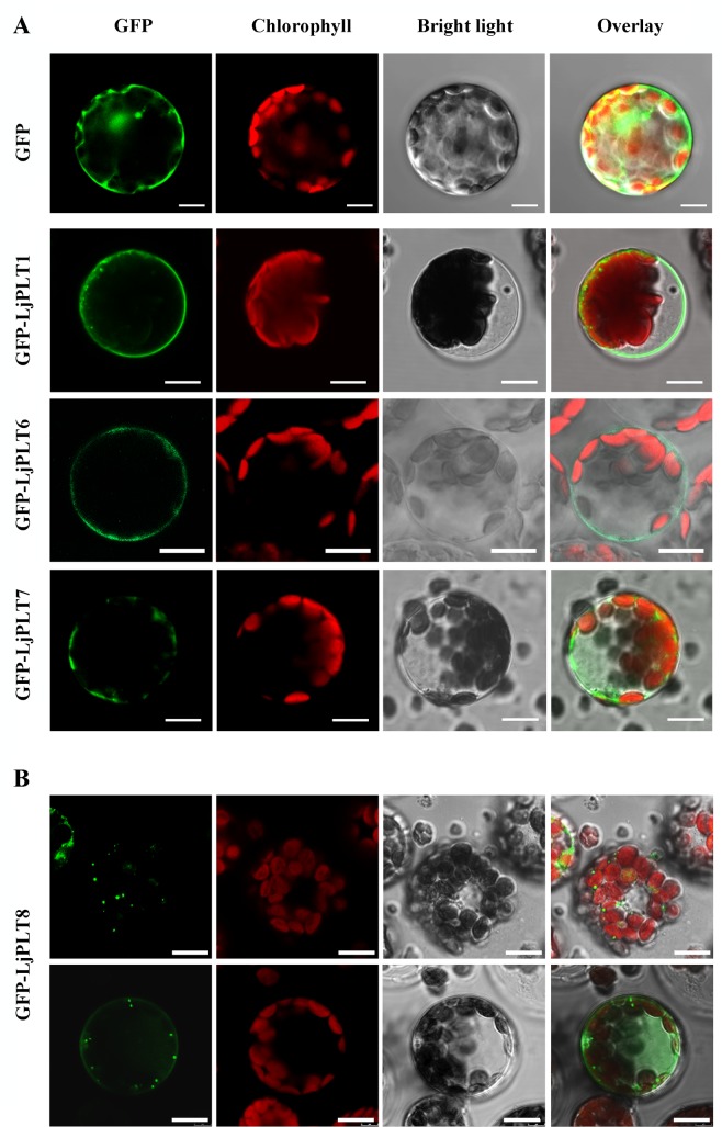
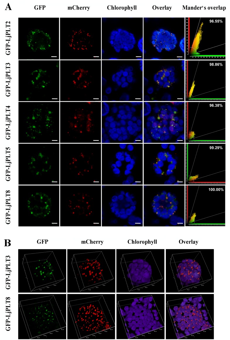
Polyols can serve as a means for the translocation of carbon skeletons and energy between source and sink organs as well as being osmoprotective solutes and antioxidants which may be involved in the resistance of some plants to biotic and abiotic stresses. Polyol/Monosaccharide transporter (PLT) proteins previously identified in plants are involved in the loading of polyols into the phloem and are reported to be located in the plasma membrane. The functions of PLT proteins in leguminous plants are not yet clear. In this study, a total of 14 putative PLT genes (LjPLT1-14) were identified in the genome of Lotus japonicus and divided into 4 clades based on phylogenetic analysis. Different patterns of expression of LjPLT genes in various tissues were validated by qRT-PCR analysis. Four genes (LjPLT3, 4, 11, and 14) from clade II were expressed at much higher levels in nodule than in other tissues. Moreover, three of these genes (LjPLT3, 4, and 14) showed significantly increased expression in roots after inoculation with Mesorhizobium loti. Three genes (LjPLT1, 3, and 9) responded when salinity and/or osmotic stresses were applied to L. japonicus. Transient expression of GFP-LjPLT fusion constructs in Arabidopsis and Nicotiana benthamiana protoplasts indicated that the LjPLT1, LjPLT6 and LjPLT7 proteins are localized to the plasma membrane, but LjPLT2 (clade IV), LjPLT3, 4, 5 (clade II) and LjPLT8 (clade III) proteins possibly reside in the Golgi apparatus. The results suggest that members of the LjPLT gene family may be involved in different biological processes, several of which may potentially play roles in nodulation in this nitrogen-fixing legume.
Conflict of interest statement
Figures
Similar articles
-
Two distinct EIN2 genes cooperatively regulate ethylene signaling in Lotus japonicus.Plant Cell Physiol. 2013 Sep;54(9):1469-77. doi: 10.1093/pcp/pct095. Epub 2013 Jul 2. Plant Cell Physiol. 2013. PMID: 23825220
-
Lotus japonicus clathrin heavy Chain1 is associated with Rho-Like GTPase ROP6 and involved in nodule formation.Plant Physiol. 2015 Apr;167(4):1497-510. doi: 10.1104/pp.114.256107. Epub 2015 Feb 25. Plant Physiol. 2015. PMID: 25717037 Free PMC article.
-
Functional characterization of polyol/monosaccharide transporter 1 in Lotus japonicus.J Plant Physiol. 2024 Jan;292:154146. doi: 10.1016/j.jplph.2023.154146. Epub 2023 Nov 24. J Plant Physiol. 2024. PMID: 38043244
-
Lotus japonicus LjKUP is induced late during nodule development and encodes a potassium transporter of the plasma membrane.Mol Plant Microbe Interact. 2004 Jul;17(7):789-97. doi: 10.1094/MPMI.2004.17.7.789. Mol Plant Microbe Interact. 2004. PMID: 15242173
-
Monosaccharide transporters in plants: structure, function and physiology.Biochim Biophys Acta. 2000 May 1;1465(1-2):263-74. doi: 10.1016/s0005-2736(00)00143-7. Biochim Biophys Acta. 2000. PMID: 10748259 Review.
Cited by
-
Micro-Evolution Analysis Reveals Diverged Patterns of Polyol Transporters in Seven Gramineae Crops.Front Genet. 2020 Jun 19;11:565. doi: 10.3389/fgene.2020.00565. eCollection 2020. Front Genet. 2020. PMID: 32636871 Free PMC article.
-
Season Affects Yield and Metabolic Profiles of Rice (Oryza sativa) under High Night Temperature Stress in the Field.Int J Mol Sci. 2020 Apr 30;21(9):3187. doi: 10.3390/ijms21093187. Int J Mol Sci. 2020. PMID: 32366031 Free PMC article.
-
Overexpression of LjPLT3 Enhances Salt Tolerance in Lotus japonicus.Int J Mol Sci. 2023 Mar 8;24(6):5149. doi: 10.3390/ijms24065149. Int J Mol Sci. 2023. PMID: 36982224 Free PMC article.
-
Roles of AGD2a in Plant Development and Microbial Interactions of Lotus japonicus.Int J Mol Sci. 2022 Jun 20;23(12):6863. doi: 10.3390/ijms23126863. Int J Mol Sci. 2022. PMID: 35743304 Free PMC article.
-
Dynamic Expression, Differential Regulation and Functional Diversity of the CNGC Family Genes in Cotton.Int J Mol Sci. 2022 Feb 12;23(4):2041. doi: 10.3390/ijms23042041. Int J Mol Sci. 2022. PMID: 35216157 Free PMC article.
References
-
- Noiraud N, Maurousset L, Lemoine R. Transport of polyols in higher plants. Plant Physiol Bioch. 2001; 39(9):717–728.
-
- Bieleski RL, Redgwell RJ. Sorbitol versus sucrose as photosynthesis and translocation products in developing apricot leaves. Aust J Plant Physiol. 1985; 12(6):657–668.
-
- Daie J. Kinetics of sugar-transport in isolated vascular bundles and phloem tissue of celery. J Am.Soc. Hortic. Sci. 1986; 111(2):216–220.
MeSH terms
Substances
LinkOut - more resources
Full Text Sources
Other Literature Sources
