. 2017 Sep 20;12(9):e0184907.
doi: 10.1371/journal.pone.0184907. eCollection 2017.

ERK1/2 signalling protects against apoptosis following endoplasmic reticulum stress but cannot provide long-term protection against BAX/BAK-independent cell death

Affiliations

ERK1/2 signalling protects against apoptosis following endoplasmic reticulum stress but cannot provide long-term protection against BAX/BAK-independent cell death

Nicola J Darling et al. PLoS One. .

Abstract

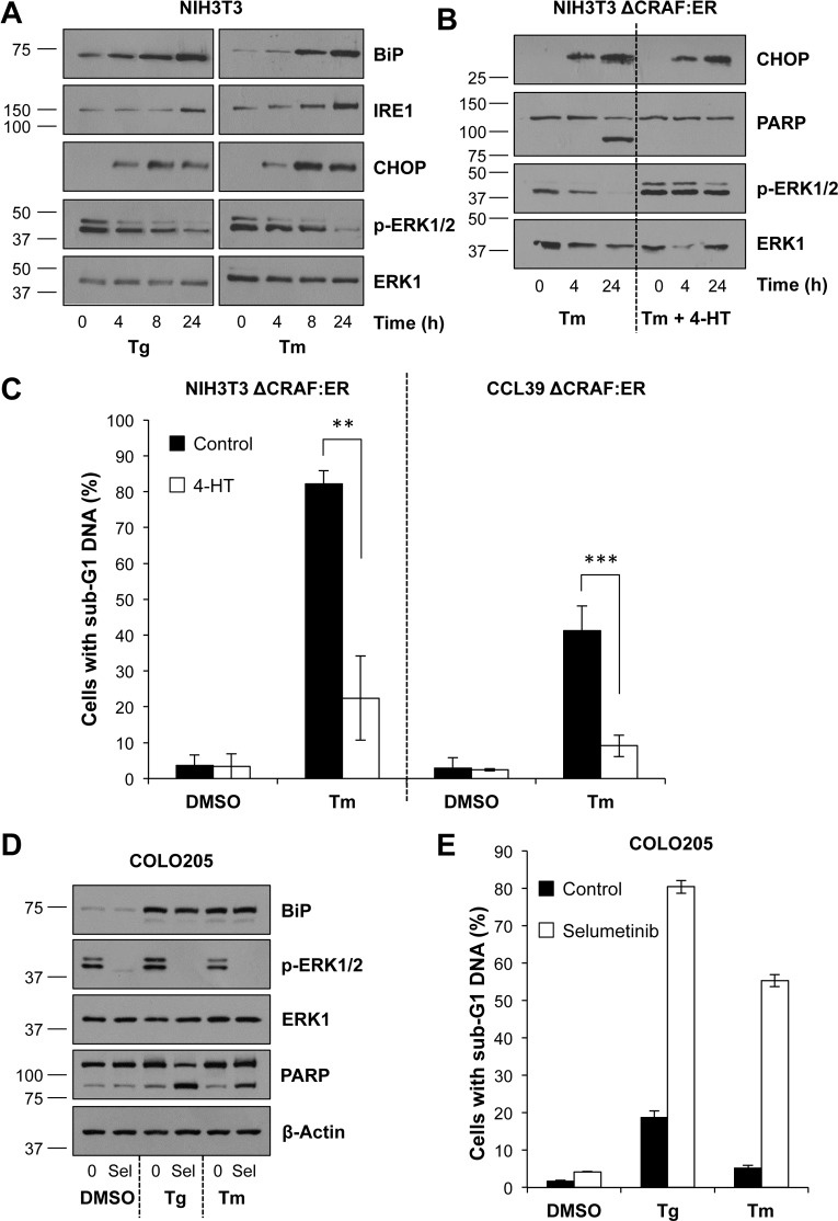
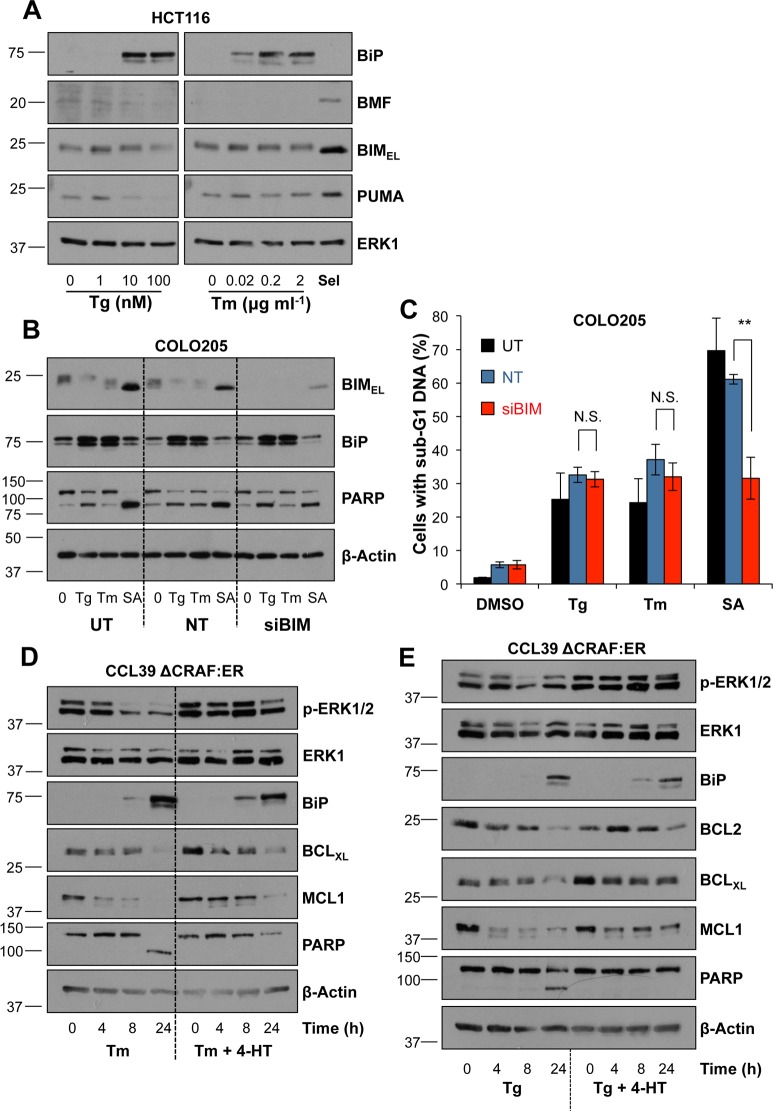
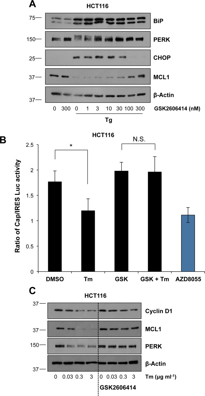
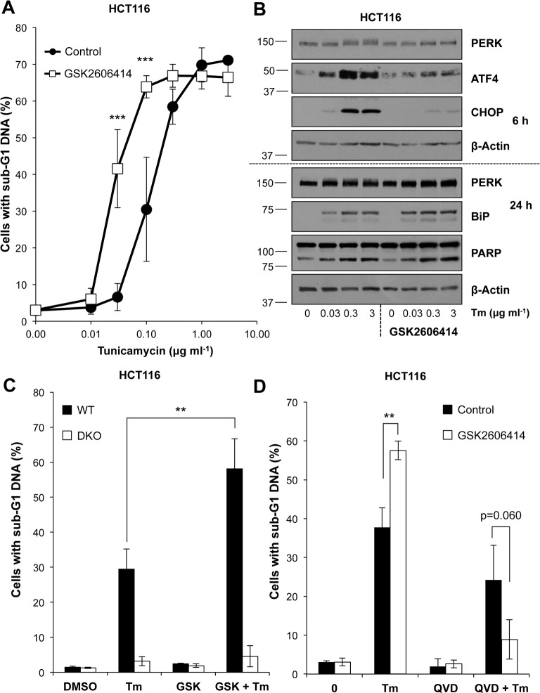
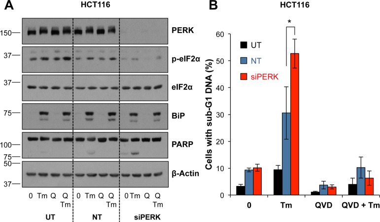
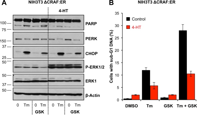
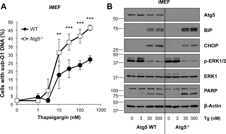
Disruption of protein folding in the endoplasmic reticulum (ER) causes ER stress. Activation of the unfolded protein response (UPR) acts to restore protein homeostasis or, if ER stress is severe or persistent, drive apoptosis, which is thought to proceed through the cell intrinsic, mitochondrial pathway. Indeed, cells that lack the key executioner proteins BAX and BAK are protected from ER stress-induced apoptosis. Here we show that chronic ER stress causes the progressive inhibition of the extracellular signal-regulated kinase (ERK1/2) signalling pathway. This is causally related to ER stress since reactivation of ERK1/2 can protect cells from ER stress-induced apoptosis whilst ERK1/2 pathway inhibition sensitises cells to ER stress. Furthermore, cancer cell lines harbouring constitutively active BRAFV600E are addicted to ERK1/2 signalling for protection against ER stress-induced cell death. ERK1/2 signalling normally represses the pro-death proteins BIM, BMF and PUMA and it has been proposed that ER stress induces BIM-dependent cell death. We found no evidence that ER stress increased the expression of these proteins; furthermore, BIM was not required for ER stress-induced death. Rather, ER stress caused the PERK-dependent inhibition of cap-dependent mRNA translation and the progressive loss of pro-survival proteins including BCL2, BCLXL and MCL1. Despite these observations, neither ERK1/2 activation nor loss of BAX/BAK could confer long-term clonogenic survival to cells exposed to ER stress. Thus, ER stress induces cell death by at least two biochemically and genetically distinct pathways: a classical BAX/BAK-dependent apoptotic response that can be inhibited by ERK1/2 signalling and an alternative ERK1/2- and BAX/BAK-independent cell death pathway.


Conflict of interest statement

Competing Interests: The authors have declared that no competing interests exist.

Figures

Fig 1
Fig 1. Activation of ERK1/2 protects against ER stress-induced cell death.
(A) NIH3T3 cells were treated with either 100 nM Tg (left panel) or 2 μg ml-1 Tm (right panel) for the indicated time. Whole cell lysates were separated by SDS-PAGE and analysed by immunoblotting using the indicated antibodies. (B) NIH3T3 ΔCRAF:ER cells were pre-treated for 1 h with 100 nM 4-HT prior to treatment with 2 μg ml-1 Tm for the indicated time. Lysates were fractionated by SDS-PAGE and analysed by immunoblotting with the indicated antibodies. Results in (A) and (B) are representative of 3 independent experiments. (C) NIH3T3 ΔCRAF:ER (left panel) or CCL39 ΔCRAF:ER (right panel) cells were pre-treated for 1 h with 100 nM 4-HT before addition of 2 μg ml-1 Tm for 48 h. Cells were then fixed, stained with propidium iodide and the proportion of cells with sub-G1 DNA was measured by flow cytometry. Results are the means ± S.D. of 3 independent experiments each performed in technical triplicate. Statistics represent the results of Student’s unpaired t-tests; **, p < 0.01; ***, p < 0.001. (D) COLO205 cells were treated for 24 h with 1 μM Selumetinib (Sel) in addition to DMSO, 30 nM Tg or 0.5 μg ml-1 Tm. Whole cell lysates were fractionated by SDS-PAGE and analysed by western blotting with the indicated antibodies. Results are representative of at least 3 independent experiments. (E) COLO205 cells were treated for 48 h as in (D). Cells were fixed, stained with propidium iodide and cell cycle distribution was determined by flow cytometry. Results are the means ± S.D. of an experiment performed in technical triplicate and representative of 3 independent experiments.
Fig 2
Fig 2. ER stress induces BAK/BAX-dependent, apoptotic cell death.
(A) HCT116 cells were treated with 100 nM Tg (top panel) or 2 μg ml-1 Tm (bottom panel) for the indicated time, whole cell lysates were fractionated by SDS-PAGE and analysed by immunoblotting with the indicated antibodies. Results are representative of at least 3 independent experiments. (B) HCT116 cells were treated with 100 nM Tg in the presence or absence of 10 μM QVD-oPh for 48 h, fixed, stained with propidium iodide and the proportion of cells with sub-G1 DNA was measured by flow cytometry. Results are the means ± S.D. of 3 experiments each performed in technical triplicate. Student’s unpaired t-test results are indicated as follows; ***, p < 0.001. (C) Whole cell lysates of HCT116, HCT116 BAK-/-, HCT116 BAX-/- or HCT116 BAK-/-, BAX-/- (DKO) cells were separated by SDS-PAGE and immunoblotted with the indicated antibodies to confirm their genotype. (D) HCT116, HCT116 BAK-/-, HCT116 BAX-/- or HCT116 BAK-/-, BAX-/- (DKO) cells were treated with 2 μg ml-1 Tm for 48 h, fixed, stained with propidium iodide and the proportion of cells with sub-G1 DNA was measured by flow cytometry. Results are the means ± S.D. of 3 independent experiments each performed in technical triplicate. Statistics represent the results of two-way ANOVA and Bonferroni post-tests comparing each genotype to WT; ***, p < 0.001. (E) HCT116, HCT116 BAK-/-, HCT116 BAX-/- or HCT116 BAK-/-, BAX-/- (DKO) cells were treated with 2 μg ml-1 Tm for 4 h (top panel) or 24 h (bottom panel). Whole cell lysates were separated by SDS-PAGE and analysed by immunoblotting with the indicated antibodies. Results are representative of three independent experiments.
Fig 3
Fig 3. ER stress-induced apoptosis does not require BIM but is accompanied by loss of multiple pro-survival proteins.
(A) HCT116 cells were treated with 1 μM Selumetinib (Sel) or the indicated concentration of Tg (left panel) or Tm (right panel) for 24 h, whole cell lysates were then separated by SDS-PAGE and analysed by immunoblotting with the indicated antibodies. Results are representative of 3 independent experiments. (B) COLO205 cells were transfected with non-targeting (NT) or BIM-specific (siBIM) siRNA or left untransfected (UT). At 48 h post-transfection cells were treated with 100 nM Tg, 2 μg ml-1 Tm or with 1 μM Selumetinib + 200nM ABT-263 (SA) for a further 24 h. Whole cell lysates were fractionated by SDS-PAGE and analysed by immunoblotting with the indicated antibodies. Results are representative of at least 3 independent experiments. (C) COLO205 cells were transfected and treated for 48 h as described in (B), fixed, stained with propidium iodide and the proportion of cells with sub-G1 DNA was measured by flow cytometry. Results are the means ± S.D. of 3 independent experiments each performed in technical triplicate, and statistics represent the results of Student’s unpaired t-tests; N.S., not significant; **, p < 0.01. (D & E) CCL39 ΔCRAF:ER cells were pre-treated for 1 h with 100 nM 4-HT prior to the addition of 2 μg ml-1 Tm (D) or 100 nM Tg (E) for the indicated time. Whole cell lysates were fractionated by SDS-PAGE and were transferred to immunoblot before analysis with the indicated antibodies. Results are representative of 3 independent experiments.
Fig 4
Fig 4. ER stress-induced inhibition of cap-dependent translation and loss of MCL1 is PERK-dependent.
(A) HCT116 cells were pre-treated for 1 h with the indicated concentration of GSK2606414 before addition of 100 nM Tg for 6 h. Whole cell lysates were separated by SDS-PAGE and analysed by immunoblotting using the indicated antibodies. (B) HCT116 cells were transfected with a dual luciferase reporter construct for assay of CAP/IRES-dependent translation. 24 h post-transfection, cells were pre-treated for 1 h with 100 nM GSK2606414 (GSK) before addition of 2 μg ml-1 Tm or 1 μM AZD8055 for 24 h. Results shown are the mean ± S.D. luciferase activity within the whole cell lysates of one experiment performed in technical triplicate and are representative of three independent experiments. Statistics shown are the results of Student’s unpaired t-tests; N.S., not significant; *, p < 0.05. (C) HCT116 cells were pre-treated for 1 h with 100 nM GSK2606414 prior to the addition of the indicated concentration of Tm for 24 h. Whole cell lysates were fractionated by SDS-PAGE and analysed by immunoblotting using the indicated antibodies. Results in (A) and (C) are representative of 3 independent experiments.
Fig 5
Fig 5. The PERK inhibitor GSK2606414 exacerbates ER stress-induced apoptosis.
(A) HCT116 cells were pre-treated for 1 h with 100 nM GSK2606414 prior to addition of the indicated concentration of Tm for 48 h. Cells were fixed, stained with propidium iodide and the proportion of cells with sub-G1 DNA was measured by flow cytometry. Results shown are the means ± S.D. of 3 independent experiments each performed in technical triplicate. Statistics represent the results of two-way ANOVA and Bonferroni post-tests; ***, p < 0.001. (B) HCT116 cells were treated as in (A) for 6 h (top panel) or 24 h (bottom panel). Whole cell lysates were analysed by immunoblotting using the indicated antibodies following separation by SDS-PAGE. Results shown are representative of 3 independent experiments. (C) HCT116 and HCT116 BAK-/-, BAX-/- (DKO) cells were pre-treated for 1 h with 100 nM GSK2606414 (GSK) before 48 h treatment with 0.1 μg ml-1 Tm. Cells were fixed, stained with propidium iodide and the proportion of cells with sub-G1 DNA was measured by flow cytometry. (D) HCT116 cells were pre-treated for 1 h with 100nM GSK2606414 prior to addition of 0.1 μg ml-1 Tm and 10 μM QVD-oPh (QVD), as indicated, for 48 h. Cells were fixed, stained with propidium iodide and the proportion of cells with sub-G1 DNA was measured by flow cytometry. Results shown in (C) and (D) are the means ± S.D. of 3 independent experiments performed in technical triplicate. Statistics represent the results of Student’s unpaired t-tests; **, p < 0.01.
Fig 6
Fig 6. RNAi-mediated silencing of PERK exacerbates ER stress-induced apoptosis.
(A) HCT116 cells were untransfected (UT) or transfected with non-targeting (NT) or PERK-targeting (siPERK) siRNA for 48 h prior to treatment with 0.1 μg ml-1 Tm and 10 μM QVD-oPh (Q) for 24 h. Whole cell lysates were fractionated by SDS-PAGE and analysed by immunoblotting using the indicated antibodies. Results shown are representative of three independent experiments. (B) HCT116 cells were transfected for 48 h as in (A) prior to treatment for 48 h with 0.1 μg ml-1 Tm and 10 μM QVD-oPh (QVD). Cells were fixed, stained with propidium iodide and the proportion of cells with sub-G1 DNA was measured by flow cytometry. Results are the combined means ± S.D. of three independent experiments performed in technical triplicate. Statistics represent the results of Student’s unpaired t-tests; *, p < 0.05.
Fig 7
Fig 7. Activation of ERK1/2 protects against cell death arising from PERK inhibition.
(A) NIH3T3 ΔCRAF:ER cells were pre-treated with 100 nM 4-HT for 1 h before addition of 100 nM GSK2606414 (GSK) and 0.1 μg ml-1 Tm alone or in combination as indicated for 24 h. Whole cell lysates were fractionated by SDS-PAGE and analysed by immunoblotting using the indicated antibodies. Results are representative of two independent experiments. (B) NIH3T3 ΔCRAF:ER cells were pre-treated with 100 nM 4-HT for 1 h before addition of 100 nM GSK2606414 (GSK) and 0.1 μg ml-1 Tm alone or in combination as indicated for 48 h. Cells were fixed and analysed by flow cytometry following propidium iodide staining. Results are means ± S.D. of three technical replicates from a single experiment and are representative of two independent experiments.
Fig 8
Fig 8. Atg5-/- immortalised MEFs exhibit enhanced ER stress-induced cell death.
(A) Atg5 WT and Atg5-/- iMEFs were treated with the indicated concentration of Tg for 48 h. Cells were fixed and analysed by flow cytometry following propidium iodide staining. Results are the means ± S.D. of 3 independent experiments performed in technical triplicate. Statistics represent the results of two-way ANOVA followed by Bonferroni post-tests; **, p < 0.01; ***, p < 0.001. (B) Alternatively Atg5 WT and Atg5-/- iMEFs were treated as in (A) for 24 h. Whole cell lysates were fractionated by SDS-PAGE prior to analysis by immunoblotting using the indicated antibodies. Results shown are representative of 3 independent experiments.

Similar articles

Cited by

References

    1. Ron D & Walter P. Signal integration in the endoplasmic reticulum unfolded protein response. Nat Rev Mol Cell Biol. 2011; 8: 519–529 - PubMed
    1. Walter P & Ron D. The unfolded protein response: from stress pathway to homeostatic regulation. Science. 2011; 334: 1081–1086 doi: 10.1126/science.1209038 - DOI - PubMed
    1. Gardner BM & Walter P. Unfolded proteins are Ire1-activating ligands that directly induce the unfolded protein response. Science. 2011; 333: 1891–1894 doi: 10.1126/science.1209126 - DOI - PMC - PubMed
    1. Harding HP, Zhang Y & Ron D. Protein translation and folding are coupled by an endoplasmic-reticulum-resident kinase. Nature. 1999; 397: 271–274 doi: 10.1038/16729 - DOI - PubMed
    1. Harding HP, Zhang Y, Bertolotti A, Zeng H and Ron D. Perk is essential for translational regulation and cell survival during the unfolded protein response. Mol Cell. 2000; 5: 897–904 - PubMed

MeSH terms

Substances