. 2017 Sep 20;10(1):438.
doi: 10.1186/s13071-017-2359-2.

Fasciclin-calcareous corpuscle binary complex mediated protein-protein interactions in Taenia solium metacestode

Affiliations

Fasciclin-calcareous corpuscle binary complex mediated protein-protein interactions in Taenia solium metacestode

Chun-Seob Ahn et al. Parasit Vectors. .

Abstract

Background: Neurocysticercosis (NC) caused by Taenia solium metacestode (TsM) is a serious neurological disease of global concern. Diverse bioactive molecules involved in the long-term survival of TsM might contribute to disease progression. Fasciclin (Fas) is an extracellular protein that mediates adhesion, migration and differentiation of cells by interacting with other molecules. We hypothesized that TsMFas might bind to calcareous corpuscle (CC) through its adhesive property and participate in crucial protein-protein interactions, thus contributing to the creation of a symbiotic interactome network.

Methods: Two paralogous TsMFas (TsMFas1 and TsMFas2) were isolated, and their molecular properties were characterized. The co-localization pattern of TsMFas1 and TsMFas2 with CC was determined. CC-TsMFas binary complex was generated by incubating CC with recombinant proteins (rTsMFas1 and 2). In vitro binding assay of CC-rTsMFas1 or CC-rTsMFas2 binary complex with TsM cellular proteins extracted from scolex and neck was conducted. Their binding partners were identified through proteomic analysis. Integrated protein-protein interaction networks were established.

Results: TsMFas1 (6072 bp long) was composed of 15 exons (841 amino acid polypeptide) interrupted by 14 introns. TsMFas2 (5201 bp long) comprised of 11 exons (597 amino acids) and 10 intervening introns. These proteins displayed 22% amino acid sequence identity to each other, but tightly conserved Fas-related domains. Several isoforms of Fas1 and Fas2 proteins might have been expressed through post-translational modifications. They showed adhesion activity with other cells. TsMFas proteins were largely distributed in parenchymal regions of the scolex and bladder wall. These molecules were co-localized with CC, a unique organelle found in platyhelminths. Subsequent proteome analysis of CC-Fas binary complex mediated protein-protein interactions revealed seven protein ligands in the TsM cellular proteins. Their functions were mainly segregated into carbohydrate metabolism (enolase, phosphoenolpyruvate carboxykinase, phosphoglycerate kinase and glyceraldehyde 3-phosphate dehydrogenase) and cytoskeleton/cellular motility (actin, paramyosin and innexin nuc-9). Those proteins had direct (physical) and/or indirect (functional) relationships along with their biochemical properties and biological roles.

Conclusion: Protein repertoires strongly suggest that TsMFas and CC may symbiotically mediate protein-protein interactions during biological processes to maintain efficacious homeostatic functions and ensure the prolonged survival of TsM in the host.

Keywords: Calcareous corpuscle; Carbohydrate metabolizing enzyme; Extracellular matrix; Fasciclin; Neurocysticercosis; Protein-protein interactions; Taenia solium metacestode.


Conflict of interest statement

Ethics approval and consent to participate

Study protocols involving the collection of adult worms from patients and T. solium metacestode from naturally infected pigs were approved by the Institutional Review Committee of Guangxi Center for Disease Prevention and Control (Protocol no. 2010–7-21). Consent was obtained in written form from patients or verbally in cases of illiterate patients. Immunization protocol was approved by the Institutional Review Board of Sungkyunkwan University (Protocol no. 2016–4). All animals were housed in AAALAC guided Institutional Animal Care and Use Committee of Sungkyunkwan University School of Medicine.

Consent for publication

Not applicable.

Competing interests

The authors declare that they have no competing interests.

Publisher’s Note

Springer Nature remains neutral with regard to jurisdictional claims in published maps and institutional affiliations.

Figures
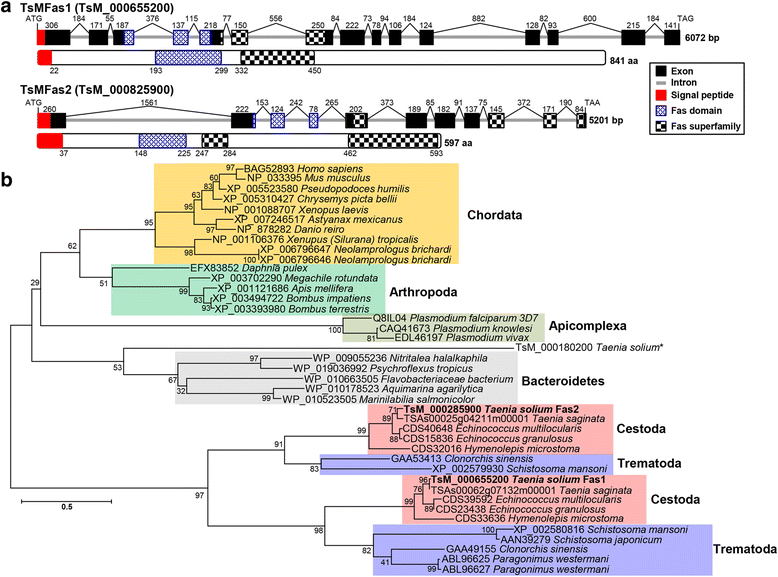
Fig. 1
Fig. 1
Molecular characteristics of Taenia solium metacestode fasciclin (TsMFas) genes. a Schematic representation of exon-intron boundary structure of TsMFas1 (TsM_000655200) and TsMFas2 (TsM_000825900) (upper panel) and domain organization of their protein products (lower panel). Exons and introns are marked by black-boxes and gray-solid lines. Numerals on the top indicate exon and intron lengths in bp. Blue and black checkered-boxes matched with fasciclin 1 and fasciclin-superfamily domains, respectively. Red boxes demonstrate signal peptides. b The phylogenetic relationships of TsMFas proteins with other 39 fasciclin-domain containing proteins. Maximum-likelihood algorithm (MEGA 6.0) was used for tree construction using full-length amino acid sequences of selected proteins. Numerals at major branching nodes indicate the percentage of appearance in 1000 bootstrap replicates. TsMFas1 and TsMFas2 proteins are highlighted in bold-face. Another fasciclin 1 found in T. solium GeneDB (TsM_000180200) is indicated by an asterisk
Fig. 2
Fig. 2
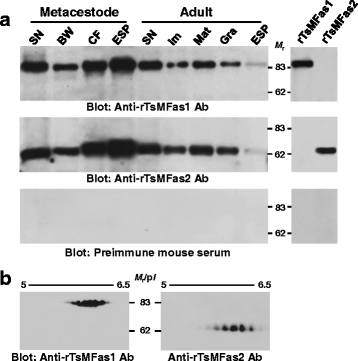
Spatiotemporal expression profile of T. solium metacestode fasciclin (TsMFas1 and TsMFas2) proteins. a Compartmental expression patterns of TsMFas1 and 2 in T. solium metacestode and adult worms. Proteins extracted from individual anatomical compartments and excretory-secretory products (each 10 μg) and recombinant proteins (each 150 ng) were separated by 8% reducing SDS-PAGE, transferred to nitrocellulose membrane and probed with anti-rTsMFas1 or anti-rTsMFas2 antibody, together with preimmune mouse serum (1:2000 dilution). The signal was detected by ECL after 2 min exposure. Lane SN: scolex/neck; Lane BW: bladder wall; Lane CF: cyst fluid; Lane ESP: excretory-secretory products; Lane Im: immature proglottid; Lane Mat: mature proglottid; Lane Gra, gravid proglottid. b Expression of different isoforms of TsMFas1 and TsMFas2. SN proteins (80 μg) were isoelectrically focused on IPG strips (pH 4–7, 7 cm long), electrophoresed by 8% SDS-PAGE and electroblotted onto a nitrocellulose membrane. Blots were probed with anti-rTsMFas1 or anti-rTsMFas2 antibody (1:2000 dilution). Immunoreactive signals were developed using ECL after 2 min exposure. Abbreviations: M r, molecular weight in kDa; pI, isoelectric point
Fig. 3
Fig. 3
Immunohistochemical localization of Fas1 and Fas2 proteins in T. solium metacestode (TsM). a, b TsM sections (4 μm thick) were incubated with anti-rTsMFas1 or anti-rTsMFas2 antibody (1:200 dilution) and further incubated with FITC-conjugated anti-mouse IgG antibody (1:500 dilution). Areas marked by a, b and c are also shown in highlight views. White arrowheads show positive reactions to calcareous corpuscles (CC). Abbreviations: BW, bladder wall; CA, spiral canal; CF, cyst fluid; SC, scolex. Scale-bars: 200 μm. c TsM sections were probed with control IgG isolated from preimmune mouse serum (1:200 dilution) and subsequently with FITC-conjugated anti-mouse IgG antibody (1:500 dilution). Markings are the same as described in a. d In vitro binding of rTsMFas1 or rTsMFas2 with CC. rTsMFas 1 or 2 protein (each 10 μg) was incubated with CC (10 μl). The binding complex was precipitated, resuspended in 2× reducing sample buffer and separated by 8% SDS-PAGE. Proteins were transblotted to nitrocellulose membranes and probed with respective antibodies (1:2000 dilution). Immune signals were detected by ECL after 2 min exposure. Lane SN: scolex/neck proteins (10 μg); Lane CC + rTsMFas1 or 2: purified CC (each 10 μl) was incubated with rTsMFas1 or 2 (10 μg each); Lane CC: calcareous corpuscles only (10 μl). Abbreviation: M r, molecular weight in kDa
Fig. 4
Fig. 4
Identification of calcareous corpuscle (CC) binding proteins of T. solium metacestode (TsM). a SDS-PAGE analysis of protein repertoire of CC. CC was purified from TsM cellular compartments using a Ficoll-Plaque gradient sedimentation. Cyst fluid (CF) and scolex/neck (SN) proteins (10 μg each) were incubated with the purified CC (10 μl). The CC-protein complex was washed with PBS, precipitated by centrifugation and resuspended in 2× SDS-PAGE sample buffer. Proteins were separated by 15% SDS-PAGE under reducing conditions. The gel was stained with CBB. Binding partners (marked by 1–16) were subjected to protein identification by LC-ESI-MS/MS. Identified proteins are listed in Additional file 3: Table S1. Lane CF: cyst fluid (10 μg) only; Lane CC + CF: CC (10 μl) was incubated with CF (10 μg); Lane CC: calcareous corpuscle only (10 μl); Lane SN: scolex/neck protein only (10 μg); Lane CC + SN: CC (10 μl) was incubated with SN protein (10 μg). Abbreviation: M r. molecular weight in kDa. Functional categorization of identified proteins from CF (b) or scolex/neck (c). Gene ontology terms assigned to the biological process, molecular function and cellular component were analyzed by Blast2GO on the basis of similarity pattern employing the second-level of GO hierarchy [28]. The number of identified proteins in each functional group is shown in histogram
Fig. 5
Fig. 5
Identification of T. solium metacestode (TsM) cellular proteins bound to calcareous corpuscle (CC)-fasciclin (TsMFas1 or TsMFas2) binary complex. a Immunoblot analysis of scolex/neck (SN) proteins depleted of TsMFas1/2 proteins. SN proteins were incubated with protein G-coupled anti-rTsMFas1/2 antibodies, and unbound proteins were eluted in flow-through fractions. Proteins (10 μg) were separated by 8% SDS-PAGE under reducing conditions and transblotted to a nitrocellulose membrane. Blots were probed with anti-rTsMFas1 or anti-rTsMFas2 antibody (1:2000 dilution). Immunoreactive signals were developed using ECL after 2 min exposure. Lane SN: whole SN proteins; Lane SNFas1/2-: SN protein depleted of Fas1/2. Abbreviation: M r., molecular weight in kDa. b TsM SN proteins depleted of Fas1 and Fas2 proteins (10 μg) were incubated with CC-rTsMFas1 or CC-rTsMFas2 binary complex (each 10 μl) and precipitated by centrifugation. Pellets were resuspended in 2× SDS-PAGE reducing sample buffer, separated on 15% gels and stained with CBB. Lane SN: scolex/neck protein (10 μg); Lane SNFas1/2-: SN protein depleted of Fas1/2 (10 μg); Lane CC: purified calcareous corpuscle (10 μl) only; Lane CC + rFas1 or 2: purified CC was incubated with rFas1 or rFas2 protein; Lane CC + SNFas1/2-: Fas1/2 depleted SN proteins were incubated with CC; Lane CC + rFas1/2 + SNFas1/2-: CC-rFas1 or CC-rFas2 binary complex was incubated with Fas1 and Fas2 depleted SN proteins. Abbreviation: M r., molecular weight in kDa. c Identification of protein repertoire for CC-Fas complex. Each protein band (7–14 and A-C) was processed for protein identification by LC-ESI-MS/MS. Independent duplicated biological samples were analysed. d Construction of protein-protein interaction network mediated by CC-Fas1 or CC-Fas2 complex by STRING algorithm ver10.0 (http://string-db.org/). Correlated interactions extracted from the platyhelminth proteins are presented with their predicted functional partners

Similar articles

Cited by

  • Comparative Analysis of Proteins of Functionally Different Body Parts of the Fish Parasites Triaenophorus nodulosus and Triaenophorus crassus.
    Borvinskaya E, Kochneva A, Bedulina D, Sukhovskaya I, Smirnov L, Babkina I. Borvinskaya E, et al. Acta Parasitol. 2021 Dec;66(4):1137-1150. doi: 10.1007/s11686-021-00384-6. Epub 2021 Apr 5. Acta Parasitol. 2021. PMID: 33818717
  • Targeted and non-targeted proteomics to characterize the parasite proteins of Echinococcus multilocularis metacestodes.
    Müller J, Preza M, Kaethner M, Rufener R, Braga S, Uldry AC, Heller M, Lundström-Stadelmann B. Müller J, et al. Front Cell Infect Microbiol. 2023 May 30;13:1170763. doi: 10.3389/fcimb.2023.1170763. eCollection 2023. Front Cell Infect Microbiol. 2023. PMID: 37325510 Free PMC article.
  • Guidelines for the use and interpretation of assays for monitoring autophagy (4th edition)1.
    Klionsky DJ, Abdel-Aziz AK, Abdelfatah S, Abdellatif M, Abdoli A, Abel S, Abeliovich H, Abildgaard MH, Abudu YP, Acevedo-Arozena A, Adamopoulos IE, Adeli K, Adolph TE, Adornetto A, Aflaki E, Agam G, Agarwal A, Aggarwal BB, Agnello M, Agostinis P, Agrewala JN, Agrotis A, Aguilar PV, Ahmad ST, Ahmed ZM, Ahumada-Castro U, Aits S, Aizawa S, Akkoc Y, Akoumianaki T, Akpinar HA, Al-Abd AM, Al-Akra L, Al-Gharaibeh A, Alaoui-Jamali MA, Alberti S, Alcocer-Gómez E, Alessandri C, Ali M, Alim Al-Bari MA, Aliwaini S, Alizadeh J, Almacellas E, Almasan A, Alonso A, Alonso GD, Altan-Bonnet N, Altieri DC, Álvarez ÉMC, Alves S, Alves da Costa C, Alzaharna MM, Amadio M, Amantini C, Amaral C, Ambrosio S, Amer AO, Ammanathan V, An Z, Andersen SU, Andrabi SA, Andrade-Silva M, Andres AM, Angelini S, Ann D, Anozie UC, Ansari MY, Antas P, Antebi A, Antón Z, Anwar T, Apetoh L, Apostolova N, Araki T, Araki Y, Arasaki K, Araújo WL, Araya J, Arden C, Arévalo MA, Arguelles S, Arias E, Arikkath J, Arimoto H, Ariosa AR, Armstrong-James D, Arnauné-Pelloquin L, Aroca A, Arroyo DS, Arsov I, Artero R, Asaro DML, Aschner M, Ashrafizadeh M, Ashur-Fabian O, Atanasov AG, Au AK, Auberger P, Auner HW, Aurelian L, Autelli R… See abstract for full author list ➔ Klionsky DJ, et al. Autophagy. 2021 Jan;17(1):1-382. doi: 10.1080/15548627.2020.1797280. Epub 2021 Feb 8. Autophagy. 2021. PMID: 33634751 Free PMC article.
  • Advances in Serological Diagnosis of Taenia solium Neurocysticercosis in Korea.
    Ahn CS, Kim JG, Huh S, Kang I, Kong Y. Ahn CS, et al. Genomics Inform. 2019 Mar;17(1):e7. doi: 10.5808/GI.2019.17.1.e7. Epub 2019 Mar 31. Genomics Inform. 2019. PMID: 30929408 Free PMC article.
  • Evolutionary Adaptations of Parasitic Flatworms to Different Oxygen Tensions.
    Martínez-González JJ, Guevara-Flores A, Del Arenal Mena IP. Martínez-González JJ, et al. Antioxidants (Basel). 2022 May 31;11(6):1102. doi: 10.3390/antiox11061102. Antioxidants (Basel). 2022. PMID: 35739999 Free PMC article. Review.

References

    1. Willingham AL, 3rd, Engels D. Control of Taenia solium cysticercosis/taeniosis. Adv Parasitol. 2006;61:509–566. doi: 10.1016/S0065-308X(05)61012-3. - DOI - PubMed
    1. John CC, Carabin H, Montano SM, Bangirana P, Zunt JR, Peterson PK. Global research priorities for infections that affect the nervous system. Nature. 2015;527:S178–S186. doi: 10.1038/nature16033. - DOI - PMC - PubMed
    1. Rajshekhara V, Joshib DD, Nguyen QD, Nguyen D, Zhou X. Taenia solium taeniosis/cysticercosis in Asia: epidemiology, impact and issues. Acta Trop. 2003;87:53–60. doi: 10.1016/S0001-706X(03)00055-X. - DOI - PubMed
    1. Engels D, Urbani C, Belotto A, Meslin F, Savioli L. The control of human (neuro)cysticercosis: which way forward? Acta Trop. 2003;87:177–182. doi: 10.1016/S0001-706X(03)00064-0. - DOI - PubMed
    1. Chung JY, Bahk YY, Huh S, Kang SY, Kong Y, Cho SY. A recombinant 10-kDa protein of Taenia solium metacestodes specific to active neurocysticercosis. J Infect Dis. 1999;180:1307–1315. doi: 10.1086/315020. - DOI - PubMed

Substances