. 2017 Nov 10;292(45):18618-18627.
doi: 10.1074/jbc.M117.810382. Epub 2017 Sep 20.

Structural basis for clonal diversity of the human T-cell response to a dominant influenza virus epitope

Affiliations

Structural basis for clonal diversity of the human T-cell response to a dominant influenza virus epitope

Xinbo Yang et al. J Biol Chem. .

Abstract

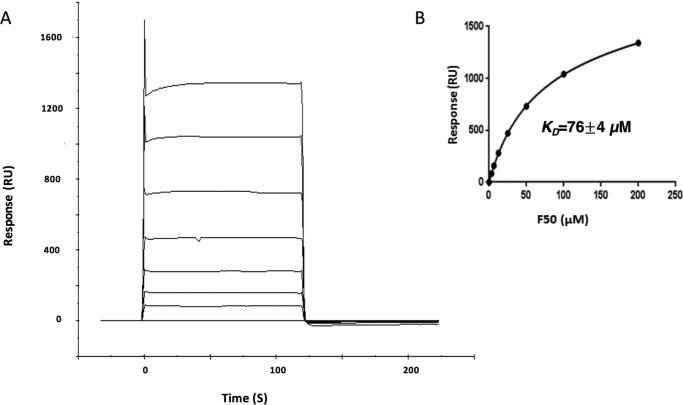
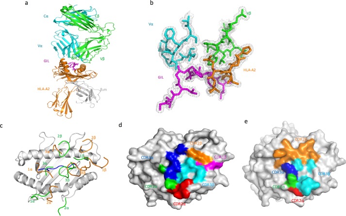
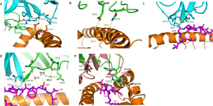
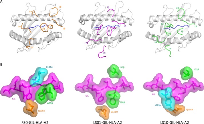
Influenza A virus (IAV) causes an acute infection in humans that is normally eliminated by CD8+ cytotoxic T lymphocytes. Individuals expressing the MHC class I molecule HLA-A2 produce cytotoxic T lymphocytes bearing T-cell receptors (TCRs) that recognize the immunodominant IAV epitope GILGFVFTL (GIL). Most GIL-specific TCRs utilize α/β chain pairs encoded by the TRAV27/TRBV19 gene combination to recognize this relatively featureless peptide epitope (canonical TCRs). However, ∼40% of GIL-specific TCRs express a wide variety of other TRAV/TRBV combinations (non-canonical TCRs). To investigate the structural underpinnings of this remarkable diversity, we determined the crystal structure of a non-canonical GIL-specific TCR (F50) expressing the TRAV13-1/TRBV27 gene combination bound to GIL-HLA-A2 to 1.7 Å resolution. Comparison of the F50-GIL-HLA-A2 complex with the previously published complex formed by a canonical TCR (JM22) revealed that F50 and JM22 engage GIL-HLA-A2 in markedly different orientations. These orientations are distinguished by crossing angles of TCR to peptide-MHC of 29° for F50 versus 69° for JM22 and by a focus by F50 on the C terminus rather than the center of the MHC α1 helix for JM22. In addition, F50, unlike JM22, uses a tryptophan instead of an arginine to fill a critical notch between GIL and the HLA-A2 α2 helix. The F50-GIL-HLA-A2 complex shows that there are multiple structurally distinct solutions to recognizing an identical peptide-MHC ligand with sufficient affinity to elicit a broad anti-IAV response that protects against viral escape and T-cell clonal loss.

Keywords: T-cell receptor; crystal structure; epitope; influenza virus; major histocompatibility complex (MHC); protein complex; surface plasmon resonance (SPR).


Conflict of interest statement

The authors declare that they have no conflicts of interest with the contents of this article

Figures

Figure 1.
Figure 1.
Surface plasmon resonance analysis of the binding of TCR F50 to GIL–HLA-A2. A, TCR F50 at concentrations of 0, 2.5, 5, 9.5, 19, 37.5, 75, and 150 μm was injected over immobilized GIL–HLA-A2 (1000 RU). B, increased RU relative to the control channel are plotted against the TCR F50 concentration. The KD (± S.D.) from fitting to a single-site equilibrium binding equation is 76 ± 4 μm for two independent experiments.
Figure 2.
Figure 2.
Structure of the TCR F50–GIL–HLA-A2 complex. a, side view of the F50–GIL–HLA-A2 complex (ribbon diagram). Cyan, TCR α chain; green, TCR β chain; orange, HLA-A2 heavy chain; gray, β2 microglobulin (βs2m); magenta, GIL peptide. b, electron density in the interface of the F50–GIL–HLA-A2 complex. Density from the final 2FoFc map at 1.7 Å resolution is contoured at 1σ. c, positions of CDR loops of TCRs F50 and JM22 (Protein Data Bank accession code 1OGA) (23) on GIL–HLA-A2. CDRs of F50 are orange. CDRs of JM22 are green. GIL peptide is blue. d, footprint of TCR F50 on GIL–HLA-A2. The top of the MHC molecule is depicted as a gray surface. The areas contacted by individual CDR loops are color-coded: green, CDR1α; red, CDR2α; blue, CDR3α; magenta, CDR1β; orange, CDR2β; cyan, CDR3β. e, footprint of TCR JM22 on GIL–HLA-A2.
Figure 3.
Figure 3.
Interactions of TCR F50 with HLA-A2 and the GIL peptide. a, interactions between CDR2α (cyan) of F50 and the HLA-A2 α2 helix (orange). The side chains of contacting residues are drawn in stick representation with carbon atoms in cyan (CDR2α) or orange (HLA-A2), nitrogen atoms in blue, and oxygen atoms in red. Hydrogen bonds are indicated by yellow dashed lines. b, interactions of CDR1β and CDR2β (green) with the HLA-A2 α1 helix (orange). c, interactions of CDR1α and CDR3α with the GIL peptide. A bridging water molecule is depicted as a red sphere. d, interactions of CDR1β and CDR3β with the GIL peptide, including bridging water molecules (red spheres). e, interactions between CDR3β (green) of F50 and GIL–HLA-A2. The side chain of βTrp99 occupies a notch between the GIL peptide (magenta) and the HLA-A2 α2 helix (orange). Superposed onto CDR3β of F50 is CDR3β of JM22 (pink). In the JM22–GIL–HLA-A2 complex (23), the pocket between GIL and the HLA-A2 α2 helix is filled by the side chain of βArg98, which makes hydrogen bonds with HLA-A2 Ala150H and Gln155H (beige).
Figure 4.
Figure 4.
Different structural solutions to binding the featureless GIL peptide. A, positions of CDR loops of TCRs F50, LS01 (Protein Data Bank accession code 5ISZ), and LS10 (Protein Data Bank accession code 5JHD) (24) on GIL–HLA-A2. CDRs of F50 are orange. CDRs of LS01 are magenta. CDRs of LS10 are green. GIL peptide is blue. LS01 and LS10 dock similarly on pMHC and differently from F50. B, top views of the F50–GIL–HLA-A2, LS01–GIL–HLA-A2, and LS10–GIL–HLA-A2 complexes in the region of the pocket between GIL (magenta) and HLA-A2 Gln155H (orange). Structures are displayed as stick/surface representations.

Similar articles

Cited by

References

    1. Zhang N., and Bevan M. J. (2011) CD8+ T cells: foot soldiers of the immune system. Immunity 35, 161–168 - PMC - PubMed
    1. Arstila T. P., Casrouge A., Baron V., Even J., Kanellopoulos J., and Kourilsky P. (1999) A direct estimate of the human αβ T cell receptor diversity. Science 286, 958–961 - PubMed
    1. Robins H. S., Srivastava S. K., Campregher P. V., Turtle C. J., Andriesen J., Riddell S. R., Carlson C. S., and Warren E. H. (2010) Overlap and effective size of the human CD8+ T cell receptor repertoire. Sci. Transl. Med. 2, 47ra64 - PMC - PubMed
    1. Qi Q., Liu Y., Cheng Y., Glanville J., Zhang D., Lee J. Y., Olshen R. A., Weyand C. M., Boyd S. D., and Goronzy J. J. (2014) Diversity and clonal selection in the human T-cell repertoire. Proc. Natl. Acad. Sci. U.S.A. 111, 13139–13144 - PMC - PubMed
    1. Li H. M., Hiroi Z., Y., Shi A., Chen G., De S., Metter E. J., Wood W. H. 3rd, Sharov A., Milner J. D., Becker K. G., Zhan M., and Weng N. P. (2016) TCRβ repertoire of CD4+ and CD8+ T cells is distinct in richness, distribution, and CDR3 amino acid composition. J. Leukoc. Biol. 99, 505–513 - PMC - PubMed

Publication types

MeSH terms