Review
. 2017 Oct;69(4):396-478.
doi: 10.1124/pr.115.012062.

Connexins in Cardiovascular and Neurovascular Health and Disease: Pharmacological Implications

Affiliations
Review

Connexins in Cardiovascular and Neurovascular Health and Disease: Pharmacological Implications

Luc Leybaert et al. Pharmacol Rev. 2017 Oct.

Abstract

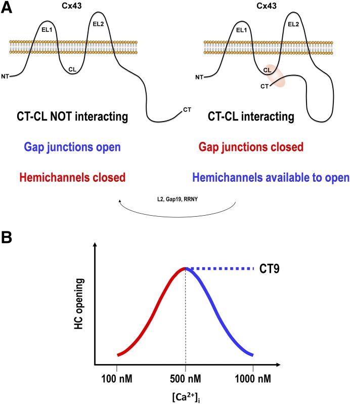
Connexins are ubiquitous channel forming proteins that assemble as plasma membrane hemichannels and as intercellular gap junction channels that directly connect cells. In the heart, gap junction channels electrically connect myocytes and specialized conductive tissues to coordinate the atrial and ventricular contraction/relaxation cycles and pump function. In blood vessels, these channels facilitate long-distance endothelial cell communication, synchronize smooth muscle cell contraction, and support endothelial-smooth muscle cell communication. In the central nervous system they form cellular syncytia and coordinate neural function. Gap junction channels are normally open and hemichannels are normally closed, but pathologic conditions may restrict gap junction communication and promote hemichannel opening, thereby disturbing a delicate cellular communication balance. Until recently, most connexin-targeting agents exhibited little specificity and several off-target effects. Recent work with peptide-based approaches has demonstrated improved specificity and opened avenues for a more rational approach toward independently modulating the function of gap junctions and hemichannels. We here review the role of connexins and their channels in cardiovascular and neurovascular health and disease, focusing on crucial regulatory aspects and identification of potential targets to modify their function. We conclude that peptide-based investigations have raised several new opportunities for interfering with connexins and their channels that may soon allow preservation of gap junction communication, inhibition of hemichannel opening, and mitigation of inflammatory signaling.


Figures

Fig. 1.
Fig. 1.
Topology of human Cx26 and Cx43 indicating crucial domains as well as peptides that affect protein and channel functions. (Left) Illustrates the extracellular loops (EL1, EL2) of Cx26 (intracellular protein parts not shown), indicating the position of highly conserved Cys residues (three per loop, yellow-filled circles) that act to stabilize the loops. Crucial residues involved in interactions between the loops upon docking of two opposed hemichannels are also indicated; for EL1, Asn-54 (green-filled circle) forms a hydrogen bond with Leu-56 of EL1 from the opposite connexin (orange), whereas Gln-57 (green) forms a hydrogen bond with Gln-57 from the opposite connexin. For EL2, salt bridges are formed between Lys-168, Asp-179, and Thr-177 from one side (green) with Asn-176 from the opposite connexin (orange). (Right) Illustrates the Cx43 topology indicating the location of conserved EL1/EL2 Cys residues (yellow). Several important domains are illustrated including the VCYD and FPISH motifs on EL1, SRPTEK on EL2 and the L2, Gap19, and CaM (calmodulin interaction site) sequences on the cytoplasmic loop (CL). Domains on the C-terminal tail (CT) include the tubulin-binding JM domain (juxtamembrane, crucial for microtubule binding), the drebrin-binding domain (Drb; crucial domain indicated; links Cx43 to F-actin), the Nedd4 domain (ubiquitin ligase), SH3 domain, and the ZO-1─binding domain (links Cx43 to F-actin). Peptides mimicking some of these domains are illustrated in the color of the corresponding domain. RRNY peptide (RRNYRRNY) is not a mimetic peptide; like L2 and Gap19, it interacts with the Cx43 CT and prevents GJ closure while inhibiting HC opening. JM2 peptide is a mouse version that differs in residue 243 from the human. The two yellow-marked CT sequences (315–326 and 340–348) are α-helical domains, whereas the rest of the CT is intrinsically disordered. The light green-marked domains are important Tyr-based sorting sequences involved in Cx43/GJ internalization (Y265AYF, Y286KLV). Blue-filled circles with white amino acid letter codes indicate mutations characterized by increased HC function relative to the function of GJs. Black-filled circles with white letter codes indicate ODDD mutations with reduced GJ and HC function. Red-filled circles with black Tyr or Ser indicate major phosphorylation sites, including CK1-targeted Ser-325,328,330, MAPK-targeted Ser-255,262,279,282, PKC-targeted Ser-368, Akt/PKB-targeted Ser-369,373, and Src/Tyk2-targeted Tyr-247,265,313. Ser-364,365 is phosphorylated by PKA but in an indirect manner involving other kinases. A detailed account on the role of the various amino acids and domains illustrated here can be found in sections II and III.
Fig. 2.
Fig. 2.
Functional effects of loop-tail interactions and [Ca2+]i elevation on Cx43 channel function. (A) Interaction of the connexin C-terminal tail (CT) with the cytoplasmic loop (CL) distinctly influences the function of gap junctions (GJs) and hemichannels (HCs). In the absence of CT-CL interaction, GJs are open while HCs are kept closed. Upon interaction of the CT with the CL, GJs are closed while HCs become available to open. The actual opening of HCs only occurs when a trigger is present, which can be of electrical (changes in membrane potential leading to depolarization or positive voltages) or chemical nature (e.g., changes in extracellular or intracellular Ca2+ concentration, inflammatory conditions, ischemic conditions including reperfusion). HC blockers like L2, Gap19 and RRNY (sequences see Fig. 1) bind to the CT and prevent CT-CL interaction, thereby driving HCs from the available to open state to the closed state. At the level of GJs, this prevents the closure of the junctional channels. (B) [Ca2+]i modulation of Cx43 HC opening. Moderate [Ca2+]i promotes HC opening via calmodulin-dependent signaling (red part of the bell-shaped curve). High [Ca2+]i inhibits HC opening by disrupting CT-CL interaction (blue part). CT9 peptide removes the high [Ca2+]i brake. For details, see sections II and III.
Fig. 3.
Fig. 3.
Chemical structure of AAP10.
Fig. 4.
Fig. 4.
Synthesis, transport, and membrane incorporation of cardiac connexins and mechanisms of ischemic damage leading to GJ closure, connexin dephosphorylation, and connexin-removal from the membrane. Molecular mechanisms and effects of antiarrhythmic peptides on GJ channels formed by Cx43 or Cx45 are also illustrated. GDP-βS, guanosine-5′-[β-thio]diphosphate trilithium salt (nonhydrolyzable GDP analog); PKCα, protein kinase Cα; CGP 54345, PKC inhibitor (inhibits only PKCα); HBDDE 2,2′,3,3′,4,4-hexahydroxy-1′-biphenyl-1-6,6′-dimethanoldimethylether, inhibitor of PKCα and PKCγ; BIM I, bisindolyl-maleimide I, inhibitor of PKCα; βI, βII, γ, δ, and ε; SR, sarcoplasmic reticulum; AAPnat, natural antiarrhythmic peptide (H-Gly-Pro-Hyp-Gly-Ala-Gly). Modified from Dhein et al. (2010).
Fig. 5.
Fig. 5.
Connexin expression in healthy arteries (A), stable atherosclerotic plaques with a thick fibrous cap (B), and after rupture of vulnerable lesions. Connexin expression is represented according to cell type. DCs, dendritic cells; ECs, endothelial cells; FC, fibrous cap; LC, lipid core MCs, monocytes; MFCs, macrophage foam cells; Plts, platelets; SMCs, smooth muscle cells; TC, T cells.
Fig. 6.
Fig. 6.
Connexin expression after myocardial ischemia (due to coronary occlusion) and reperfusion in wild-type mice. (Left) Cx37, Cx40, and Cx43 expression (in green) in unaffected healthy (H) myocardium. (Right) Cx37, Cx40, and Cx43 expression (in green) at the border zone (B) of the infarcted area (I). Tissue is counterstained with Evans Blue (in red), and nuclei are stained with DAPI (in blue).
Fig. 7.
Fig. 7.
Roles of Cx43 in cardiac ischemia-reperfusion. Gap junctions close under ischemic conditions (“healing over” caused by the low pH and elevated [Ca2+]i) but substantial coupling may persist after ischemia-reperfusion (Ruiz-Meana et al., 2001). Open GJs may act beneficially by supplying essential metabolites to neighboring cells but may also spread injury signals, causing cell death propagation (reviewed in Decrock et al., 2009b; Michela et al., 2015). GJs have been implicated in the spreading of hypercontracture necrosis in a process mediated by Na+ flux through GJs (reviewed in García-Dorado et al., 2004). Uncontrolled hemichannel (HC) opening facilitates ionic fluxes that may lead to cell swelling (Wang et al., 2013c). Cx43 is also present in mitochondria where they are involved in the signaling cascade of ischemic preconditioning, conferring cardioprotective effects (reviewed in Schulz et al., 2007; Miura et al., 2010; Schulz et al., 2015). Mitochondrial Cx43 has been demonstrated to form hexameric structures involved in inner mitochondrial membrane K+ fluxes, pointing to functional HCs (Miro-Casas et al., 2009).
Fig. 8.
Fig. 8.
Composite map of the Cx43 polypeptide depicting all the known GJA1 gene mutations linked to ODDD.
Fig. 9.
Fig. 9.
Hemichannel-linked “pathologic pore” roles in injury spread and the perpetuation of chronic disease. Hemichannel opening plays a key role in cellular edema, lesion spread, initiation, and perpetuation of both the adaptive and innate immune responses and in particular in chronic disease conditions and the initiation and perpetuation of the inflammasome pathway. Multiple pathologic signals have been shown to trigger HC opening, resulting in pathologic levels of extracellular inflammatory stimulators such as ATP (with subsequent ATP-induced ATP release) and glutamate, the onset of calcium waves and seizures, and changes in the cytoplasmic ionic composition that may trigger apoptosis or loss of a cell’s ability to osmoregulate. Loss of vascular integrity to breach the blood-brain or blood-retina barrier may be an initiating event, but conversely, inflammation leads to loss of vascular integrity, including rupture of endothelial cells and vascular dropout. The blue arrows to adjacent boxes indicate reported outcomes of HC opening in different cell types (irrespective of the specific connexin isoform expressed). Not all effects necessarily occur at once, depending upon the type and extent of injury and the specific cell type involved. These pathways have been implicated in a remarkable number of central nervous system cerebrovascular and retinovascular indications, including trauma, such as spinal cord injury; ischemia; stroke; and infectious disease and chronic diseases, including Alzheimer’s, Parkinson’s, amyotrophic lateral sclerosis, diabetic retinopathy, macular edema, age-related macular degeneration, and chronic pain. In several models, intervention using connexin HC blockers has been exploited to break the cycle, reducing the extent of damage after an acute insult or breaking the inflammatory cycle in chronic disease conditions. Red arrows at the (top) and (bottom) emphasize the HC-mediated feedback loops that contribute to damage spread and perpetuation of the inflammatory response.

Similar articles

Cited by

References

    1. Abascal F, Zardoya R. (2013) Evolutionary analyses of gap junction protein families. Biochim Biophys Acta 1828:4–14. - PubMed
    1. Abcouwer SF, Lin CM, Wolpert EB, Shanmugam S, Schaefer EW, Freeman WM, Barber AJ, Antonetti DA. (2010) Effects of ischemic preconditioning and bevacizumab on apoptosis and vascular permeability following retinal ischemia-reperfusion injury. Invest Ophthalmol Vis Sci 51:5920–5933. - PubMed
    1. Abrams CK, Scherer SS. (2012) Gap junctions in inherited human disorders of the central nervous system. Biochim Biophys Acta 1818:2030–2047. - PMC - PubMed
    1. Abudara V, Bechberger J, Freitas-Andrade M, De Bock M, Wang N, Bultynck G, Naus CC, Leybaert L, Giaume C. (2014) The connexin43 mimetic peptide Gap19 inhibits hemichannels without altering gap junctional communication in astrocytes. Front Cell Neurosci 8:306. - PMC - PubMed
    1. Agullo-Pascual E, Cerrone M, Delmar M. (2014a) Arrhythmogenic cardiomyopathy and Brugada syndrome: diseases of the connexome. FEBS Lett 588:1322–1330. - PMC - PubMed

Publication types

MeSH terms