

Predicting epiglottic collapse in patients with obstructive sleep apnoea
- PMID: 28931660
- PMCID: PMC5915305
- DOI: 10.1183/13993003.00345-2017
Predicting epiglottic collapse in patients with obstructive sleep apnoea
Abstract
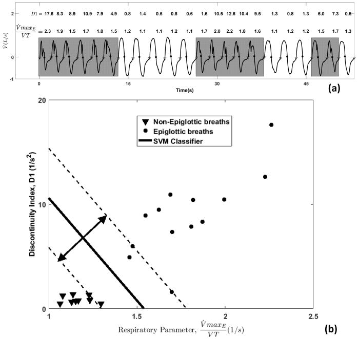
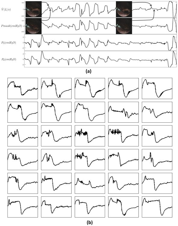
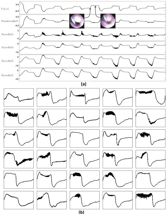
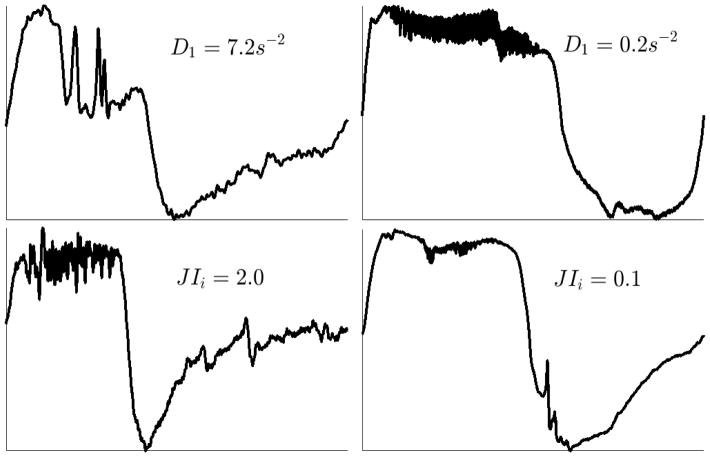
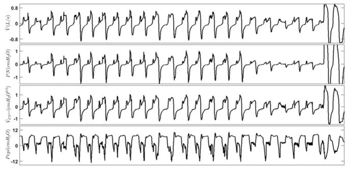
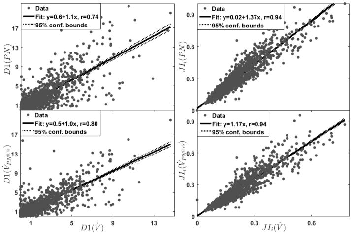
Obstructive sleep apnoea (OSA) is characterised by pharyngeal obstruction occurring at different sites. Endoscopic studies reveal that epiglottic collapse renders patients at higher risk of failed oral appliance therapy or accentuated collapse on continuous positive airway pressure. Diagnosing epiglottic collapse currently requires invasive studies (imaging and endoscopy). As an alternative, we propose that epiglottic collapse can be detected from the distinct airflow patterns it produces during sleep.23 OSA patients underwent natural sleep endoscopy. 1232 breaths were scored as epiglottic/nonepiglottic collapse. Several flow characteristics were determined from the flow signal (recorded simultaneously with endoscopy) and used to build a predictive model to distinguish epiglottic from nonepiglottic collapse. Additionally, 10 OSA patients were studied to validate the pneumotachograph flow features using nasal pressure signals.Epiglottic collapse was characterised by a rapid fall(s) in the inspiratory flow, more variable inspiratory and expiratory flow and reduced tidal volume. The cross-validated accuracy was 84%. Predictive features obtained from pneumotachograph flow and nasal pressure were strongly correlated.This study demonstrates that epiglottic collapse can be identified from the airflow signal measured during a sleep study. This method may enable clinicians to use clinically collected data to characterise underlying physiology and improve treatment decisions.
Copyright ©ERS 2017.
Conflict of interest statement
Conflict of interest: Disclosures can be found alongside this article at erj.ersjournals.com
Figures
Comment in
-
Two valves in the pharynx.Eur Respir J. 2017 Sep 20;50(3):1701496. doi: 10.1183/13993003.01496-2017. Print 2017 Sep. Eur Respir J. 2017. PMID: 28931669 No abstract available.
Similar articles
-
Home sleep study nasal airflow patterns and epiglottic collapse during drug induced sleep endoscopy.Sleep Breath. 2025 Mar 28;29(2):142. doi: 10.1007/s11325-025-03305-3. Sleep Breath. 2025. PMID: 40153093
-
Effect of Sleeping Position on Upper Airway Patency in Obstructive Sleep Apnea Is Determined by the Pharyngeal Structure Causing Collapse.Sleep. 2017 Mar 1;40(3):zsx005. doi: 10.1093/sleep/zsx005. Sleep. 2017. PMID: 28329099 Free PMC article.
-
Palatal prolapse as a signature of expiratory flow limitation and inspiratory palatal collapse in patients with obstructive sleep apnoea.Eur Respir J. 2018 Feb 14;51(2):1701419. doi: 10.1183/13993003.01419-2017. Print 2018 Feb. Eur Respir J. 2018. PMID: 29444914 Free PMC article.
-
[Research progress of obstructive sleep apnea syndrome caused by epiglottic collapse].Lin Chuang Er Bi Yan Hou Tou Jing Wai Ke Za Zhi. 2019 Feb 5;33(2):186-189. doi: 10.13201/j.issn.1001-1781.2019.02.024. Lin Chuang Er Bi Yan Hou Tou Jing Wai Ke Za Zhi. 2019. PMID: 30808152 Review. Chinese.
-
Surgical Treatment Options for Epiglottic Collapse in Adult Obstructive Sleep Apnoea: A Systematic Review.Life (Basel). 2022 Nov 11;12(11):1845. doi: 10.3390/life12111845. Life (Basel). 2022. PMID: 36430980 Free PMC article. Review.
Cited by
-
Flow-Identified Site of Collapse During Drug-Induced Sleep Endoscopy: Feasibility and Preliminary Results.Chest. 2021 Feb;159(2):828-832. doi: 10.1016/j.chest.2020.09.079. Epub 2020 Sep 14. Chest. 2021. PMID: 32941861 Free PMC article. No abstract available.
-
Structure and severity of pharyngeal obstruction determine oral appliance efficacy in sleep apnoea.J Physiol. 2019 Nov;597(22):5399-5410. doi: 10.1113/JP278164. Epub 2019 Oct 1. J Physiol. 2019. PMID: 31503323 Free PMC article.
-
Phenotypic Labelling Using Drug-Induced Sleep Endoscopy Improves Patient Selection for Mandibular Advancement Device Outcome: A Prospective Study.J Clin Sleep Med. 2019 Aug 15;15(8):1089-1099. doi: 10.5664/jcsm.7796. J Clin Sleep Med. 2019. PMID: 31482830 Free PMC article.
-
Home sleep study nasal airflow patterns and epiglottic collapse during drug induced sleep endoscopy.Sleep Breath. 2025 Mar 28;29(2):142. doi: 10.1007/s11325-025-03305-3. Sleep Breath. 2025. PMID: 40153093
-
Patients with epiglottic collapse showed less severe obstructive sleep apnea and good response to treatment other than continuous positive airway pressure: a case-control study of 224 patients.J Clin Sleep Med. 2021 Mar 1;17(3):413-419. doi: 10.5664/jcsm.8904. J Clin Sleep Med. 2021. PMID: 33094721 Free PMC article.
References
-
- Young T, Peppard P, Gottlieb D. The epidemiology of obstructive sleep apnea: a population health perspective. Am J Respir Crit Care Med. 2002;165:1217–1239. - PubMed
-
- Colt HG, Hass H, Rich GB. Hypoxemia vs sleep fragmentation as cause of excessive daytime sleepiness in obstructive sleep apnea. CHEST Journal. 1991;100(6):1542–1548. - PubMed
-
- Kezirian EJ, Hohenhorst W, de Vries N. Drug-induced sleep endoscopy: the VOTE classification. Eur Arch Otorhinolaryngol. 2011;268(8):1233–1236. - PubMed
-
- Vroegop AV, Vanderveken OM, Boudewyns AN, Scholman J, Saldien V, Wouters K, Braem MJ, Van de Heyning PH, Hamans E. Drug-induced sleep endoscopy in sleep-disordered breathing: report on 1,249 cases. Laryngoscope. 2014;124(3):797–802. - PubMed
Publication types
MeSH terms
Grants and funding
LinkOut - more resources
Full Text Sources
Other Literature Sources
Molecular Biology Databases
Research Materials