. 2017 Nov 14;91(23):e01324-17.
doi: 10.1128/JVI.01324-17. Print 2017 Dec 1.

p21 Restricts HIV-1 in Monocyte-Derived Dendritic Cells through the Reduction of Deoxynucleoside Triphosphate Biosynthesis and Regulation of SAMHD1 Antiviral Activity

Affiliations

p21 Restricts HIV-1 in Monocyte-Derived Dendritic Cells through the Reduction of Deoxynucleoside Triphosphate Biosynthesis and Regulation of SAMHD1 Antiviral Activity

Jose Carlos Valle-Casuso et al. J Virol. .

Abstract

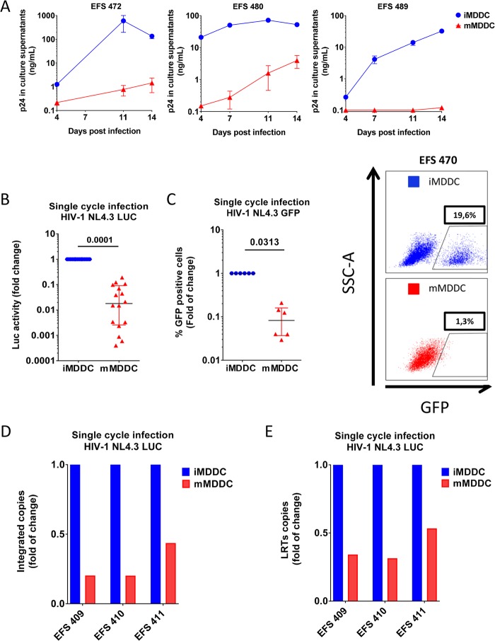
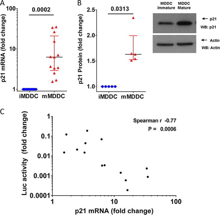
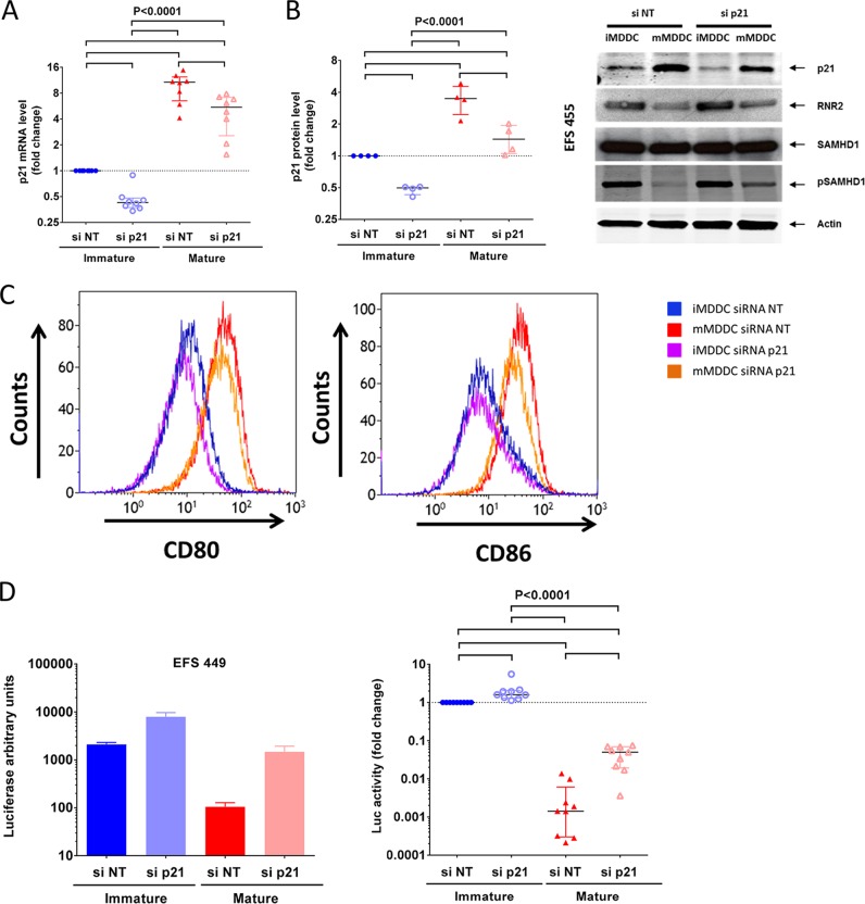
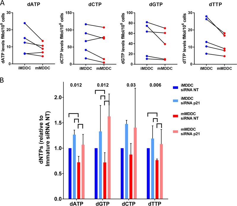
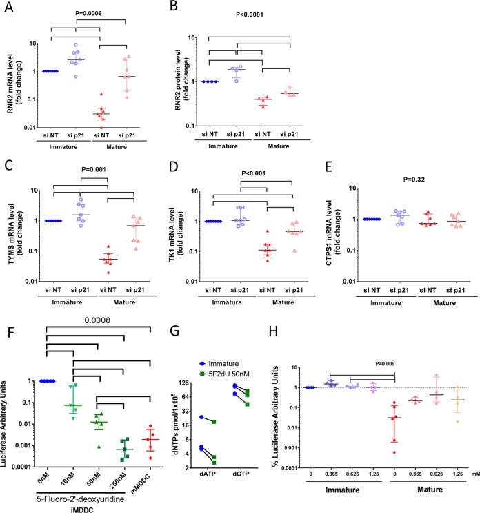
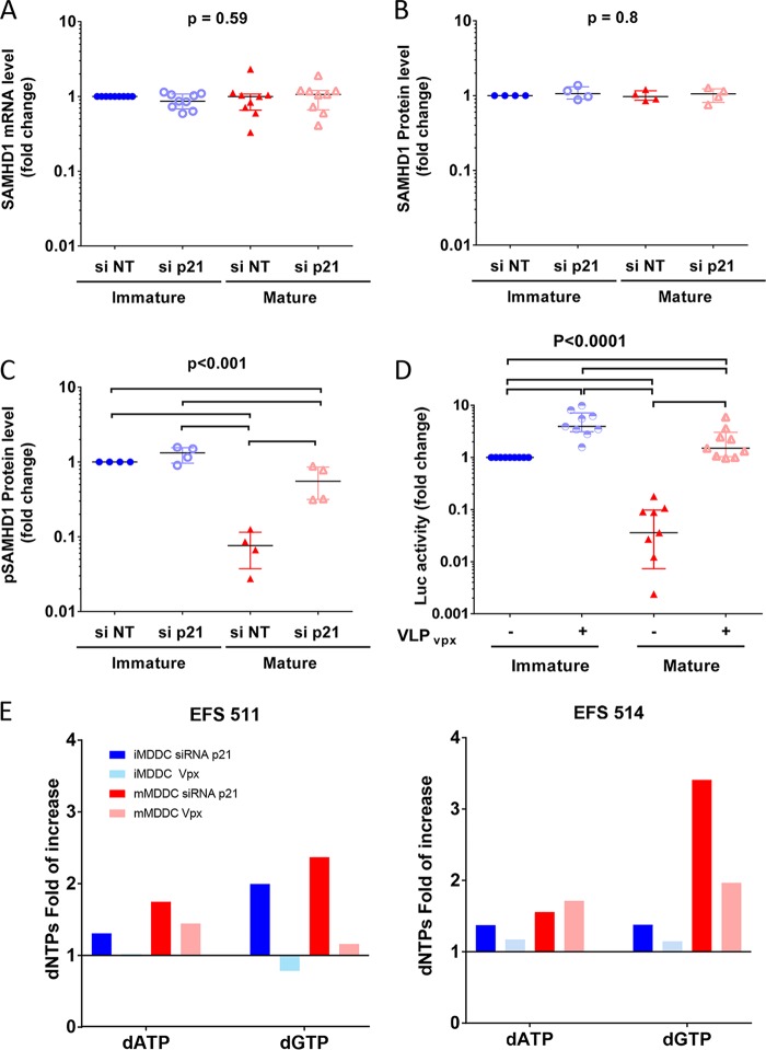
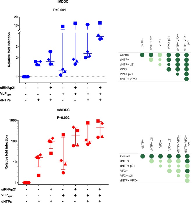
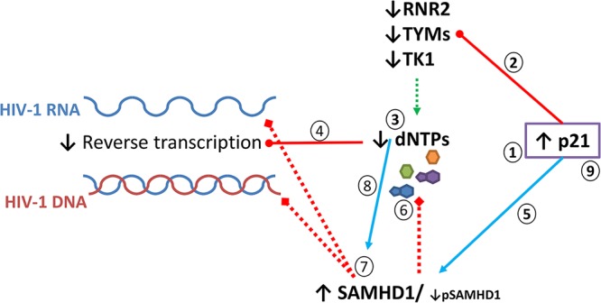
HIV-1 infection of noncycling cells, such as dendritic cells (DCs), is impaired due to limited availability of deoxynucleoside triphosphates (dNTPs), which are needed for HIV-1 reverse transcription. The levels of dNTPs are tightly regulated during the cell cycle and depend on the balance between dNTP biosynthesis and degradation. SAMHD1 potently blocks HIV-1 replication in DCs, although the underlying mechanism is still unclear. SAMHD1 has been reported to be able to degrade dNTPs and viral nucleic acids, which may both hamper HIV-1 reverse transcription. The relative contribution of these activities may differ in cycling and noncycling cells. Here, we show that inhibition of HIV-1 replication in monocyte-derived DCs (MDDCs) is associated with an increased expression of p21cip1/waf, a cell cycle regulator that is involved in the differentiation and maturation of DCs. Induction of p21 in MDDCs decreases the pool of dNTPs and increases the antiviral active isoform of SAMHD1. Although both processes are complementary in inhibiting HIV-1 replication, the antiviral activity of SAMHD1 in our primary cell model appears to be, at least partially, independent of its dNTPase activity. The reduction in the pool of dNTPs in MDDCs appears rather mostly due to a p21-mediated suppression of several enzymes involved in dNTP synthesis (i.e., RNR2, TYMS, and TK-1). These results are important to better understand the interplay between HIV-1 and DCs and may inform the design of new therapeutic approaches to decrease viral dissemination and improve immune responses against HIV-1.IMPORTANCE DCs play a key role in the induction of immune responses against HIV. However, HIV has evolved ways to exploit these cells, facilitating immune evasion and virus dissemination. We have found that the expression of p21, a cyclin-dependent kinase inhibitor involved in cell cycle regulation and monocyte differentiation and maturation, potentially can contribute to the inhibition of HIV-1 replication in monocyte-derived DCs through multiple mechanisms. p21 decreased the size of the intracellular dNTP pool. In parallel, p21 prevented SAMHD1 phosphorylation and promoted SAMHD1 dNTPase-independent antiviral activity. Thus, induction of p21 resulted in conditions that allowed the effective inhibition of HIV-1 replication through complementary mechanisms. Overall, p21 appears to be a key regulator of HIV infection in myeloid cells.

Keywords: HIV replication; HIV-1; SAMHD1; cellular factors; dNTPs; dendritic cells; p21.


Figures

FIG 1
FIG 1
Mature MDDCs have low susceptibility to HIV-1 infection, which is blocked at the reverse transcription level. (A) p24 measured in culture supernatants after the infection of immature MDDCs (iMDDCs; blue) and mature MDDCs (mMDDCs; red) with HIV-1 BaL in 3 independent experiments. The means and standard deviations for three replicates are shown at each time point. (B and C) Luciferase activity (B) or percentage of GFP+ cells (C) in mature MDDCs compared with that in immature MDDCs after infection with single-cycle HIV-1 NL4.3Δenv VSV-G particles carrying luciferase (n = 16) or GFP (n = 6) reporter genes. Each symbol represents results with cells from different donors. The median and IQR values are shown. (Right) One representative example of GFP production in immature and mature MDDCs is shown. (D and E) Relative number of integrated Alu-long terminal repeat (LTR) copies (D) or U5-Gag copies (E) quantified by RT-qPCR in mature MDDCs compared with that in immature MDDCs 72 h after infection with single-cycle HIV-1 NL4.3ΔenvLuc VSV-G particles. Viral cDNAs were normalized to the albumin gene content in each sample. Experiments with cells from three different donors are shown. Bars represent the means from 2 replicates.
FIG 2
FIG 2
p21 induction in mature MDDCs correlates with viral replication levels. (A) p21 mRNA expression was quantified by qRT-PCR in immature (blue) and mature (red) MDDCs. The data are expressed as the fold change in the number of p21 copies in mature MDDCs compared to that in immature MDDCs. Each symbol represents the results obtained with cells from one donor (n = 13). The median and IQR values are shown. (B) Analysis of protein expression in immature and mature MDDCs by Western blotting (WB). (Left) p21 protein expression quantification are expressed as the relative change in p21 protein expression in mature MDDCs compared with that in immature MDDCs in the same experiments. The median and IQR values of independent experiments (n = 5 donors) are depicted. An example of p21 protein analyzed by Western blotting is shown. (C) Correlation between relative changes in the number of p21 mRNA copies and relative changes in luciferase activity after infection with HIV-1 NL4.3ΔenvLuc VSV-G in mature MDDCs compared with those in immature MDDCs from 14 different donors.
FIG 3
FIG 3
Downregulation of p21 expression levels by siRNA in mature MDDCs restores viral replication. (A and B) The relative number of copies of p21 mRNA (A) and the relative p21 protein levels (B) in immature and mature MDDCs after transfection with siRNA targeting p21 (siRNA p21) or a pool of nontargeting siRNA (siRNA NT). The data are expressed in relation to the number of mRNA copies or protein levels in immature MDDCs transfected with nontargeting siRNA. Symbols represent experiments with cells from different donors (n = 8 for mRNA and n = 4 for protein). The median and IQR values are shown. An example of Western blot analysis of the different proteins studied is shown on the right. (C) Level of expression of CD86, CD80, and CD40 on the surface of MDDCs transfected with siRNA NT or siRNA p21 and cultured in the absence or presence of CD40L and IFN-γ. (D) Changes in luciferase activity in immature and mature MDDCs transfected with siRNA NT or siRNA p21 after infection with HIV-1 NL4.3ΔenvLuc VSV-G. The results are shown as the fold change relative to the luciferase activity in immature MDDCs transfected with siRNA NT. Symbols represent independent experiments (n = 8 donors). The median and IQR values are shown. One representative experiment is shown on the left (means and standard deviations from three replicates). P values of ANOVA are shown. Pairwise significant differences (P < 0.05) in post hoc analyses are identified by horizontal lines.
FIG 4
FIG 4
Maturation of MDDCs decreases the size of the intracellular dNTP pools, but the pool sizes recover upon p21 knockdown. (A) Intracellular levels of dATP, dCTP, dGTP, and dTTP quantified by single-nucleotide primer extension gel analysis in immature and mature MDDCs. Independent experiments with cells from 5 donors are shown. (B) Changes in the dNTP levels in immature and mature MDDCs transfected with siRNA NT or siRNA p21. The results are shown as fold change relative to each dNTP level in immature MDDCs transfected with siRNA NT. The median and IQR values of the results obtained with cells from 4 different donors are shown (n = 4 donors). P values of ANOVA are shown. Significant differences (P < 0.05) between conditions in post hoc analyses are identified by horizontal lines.
FIG 5
FIG 5
p21 regulates the de novo and salvage dNTP synthesis pathways. (A to E) Relative levels of RNR2 mRNA (A), relative RNR2 protein levels (B), relative levels of thymidylate synthase (TYMS) mRNA (C), relative levels of thymidine kinase 1 (TK-1) mRNA (D), and relative levels of CTP synthase 1 (CTPS1) mRNA (E) in immature and mature MDDCs after transfection with siRNA p21 or siRNA NT. The data are expressed relative to the number of mRNA copies or protein levels in immature MDDCs transfected with nontargeting siRNA. Symbols represent independent experiments (n = 7 donors for mRNA and n = 4 donors for RNR2 protein). (F) Changes in luciferase activity after infection with HIV-1 NL4.3ΔenvLuc VSV-G in immature MDDCs cultured in the presence of increasing amounts of 5-fluoro-2′-deoxyuridine. The results with cells from five donors are shown. (G) Changes in intracellular levels of dATP and dGTP quantified by single-nucleotide primer extension gel analysis in immature MDDCs after treatment with 50 nM 5-fluoro-2′-deoxyuridine. Independent experiments with cells from 3 donors are shown. (H) Changes in luciferase activity after infection with HIV-1 NL4.3ΔenvLuc VSV-G in immature and mature MDDCs cultured in the presence of increasing amounts of exogenous dNTPs. The results are shown as fold change relative to the luciferase activity in immature MDDCs cultured in the absence of exogenous dNTPs. The symbols represent independent experiments (n = 5 donors). The median and IQR values are shown. P values of ANOVA are shown. Significant differences (P < 0.05) between conditions in post hoc analyses are identified by horizontal lines.
FIG 6
FIG 6
p21 expression modulates SAMHD1 phosphorylation levels in MDDCs. (A to C) Relative copies of SAMHD1 mRNA (A), total SAMHD1 protein levels (B), and pSAMHD1 protein levels (C) in immature and mature MDDC after transfection with siRNA p21 or siRNA NT. The data are expressed relative to the number of mRNA copies or protein levels in immature MDDCs transfected with nontargeting siRNA. The symbols represent independent experiments (n = 9 donors for mRNA and n = 4 donors for proteins). (D) Changes in luciferase activity after infection with HIV-1 NL4.3ΔenvLuc VSV-G in immature and mature MDDCs cultured in the presence of virus-like particles with or without the SIV VPX protein. The results are shown as the fold change relative to the luciferase activity in immature MDDCs cultured with VLPs not carrying VPX. The symbols represent independent experiments (n = 9). The median and IQR values are shown. P values of ANOVA are shown. Significant differences (P < 0.05) between conditions in post hoc analyses are identified by horizontal lines. (E) Changes in the dNTP levels in immature and mature MDDCs transfected with siRNA p21 or treated with VLP-VPX particles. The results are shown as fold change in dNTP levels relative to those of control nontreated immature or mature MDDCs, respectively.
FIG 7
FIG 7
p21 controls the HIV-1 inhibition in mature MDDCs through two synergistic mechanisms. Changes in luciferase activity after infection with HIV-1 NL4.3ΔenvLuc VSV-G in immature (top) or mature (bottom) MDDCs transfected with siRNA p21 or siRNA NT, pretreated with VLPs with or without VPX, and cultured in the absence or presence of exogenous dNTPs (0.625 nM). The data are expressed as the fold change in luciferase activity relative to the control condition for immature or mature MDDCs. The median and IQR values of independent experiments with cells from 4 different donors are shown. Each donor is identified with a different symbol. Panels on the right summarize the following comparisons between the different conditions: statistically significant differences in post hoc analyses are identified by dark green circles, and big circles indicate consistent differences between the conditions tested in all 4 experiments.
FIG 8
FIG 8
Proposed model for p21-mediated HIV-1 restriction in MDDCs. The expression of p21 is induced during MDDC maturation (step 1). The increase in p21 expression leads to the downregulation of several enzymes (RNR2, TYMS, and TK1) involved in dNTP biosynthesis (step 2), which would decrease the dNTP pool size (step 3), impairing HIV-1 reverse transcription (step 4). In parallel, p21 induction could increase the active isoform of SAMHD1 (step 5), which would further block viral replication through the degradation of dNTPs (step 6) or exonuclease activity (step 7). Low levels of dNTP synthesis as a consequence of p21 induction might further facilitate the exonuclease activity of SAMHD1 (step 8). Although not explored here, induction of p21 might also directly block reverse transcriptase activity and impair HIV transcription, as has been shown in CD4+ T cells (step 9) (66, 67).

Similar articles

Cited by

References

    1. Manches O, Frleta D, Bhardwaj N. 2014. Dendritic cells in progression and pathology of HIV infection. Trends Immunol 35:114–122. doi:10.1016/j.it.2013.10.003. - DOI - PMC - PubMed
    1. Burleigh L, Lozach PY, Schiffer C, Staropoli I, Pezo V, Porrot F, Canque B, Virelizier JL, Arenzana-Seisdedos F, Amara A. 2006. Infection of dendritic cells (DCs), not DC-SIGN-mediated internalization of human immunodeficiency virus, is required for long-term transfer of virus to T cells. J Virol 80:2949–2957. doi:10.1128/JVI.80.6.2949-2957.2006. - DOI - PMC - PubMed
    1. Coleman CM, Wu L. 2009. HIV interactions with monocytes and dendritic cells: viral latency and reservoirs. Retrovirology 6:51. doi:10.1186/1742-4690-6-51. - DOI - PMC - PubMed
    1. Nobile C, Petit C, Moris A, Skrabal K, Abastado JP, Mammano F, Schwartz O. 2005. Covert human immunodeficiency virus replication in dendritic cells and in DC-SIGN-expressing cells promotes long-term transmission to lymphocytes. J Virol 79:5386–5399. doi:10.1128/JVI.79.9.5386-5399.2005. - DOI - PMC - PubMed
    1. Wu L, Kewal Ramani VN. 2006. Dendritic-cell interactions with HIV: infection and viral dissemination. Nat Rev Immunol 6:859–868. doi:10.1038/nri1960. - DOI - PMC - PubMed

Publication types

MeSH terms