. 2017 Nov 14;91(23):e01340-17.
doi: 10.1128/JVI.01340-17. Print 2017 Dec 1.

A DNA Vaccine That Targets Hemagglutinin to Antigen-Presenting Cells Protects Mice against H7 Influenza

Affiliations

A DNA Vaccine That Targets Hemagglutinin to Antigen-Presenting Cells Protects Mice against H7 Influenza

Tor Kristian Andersen et al. J Virol. .

Abstract

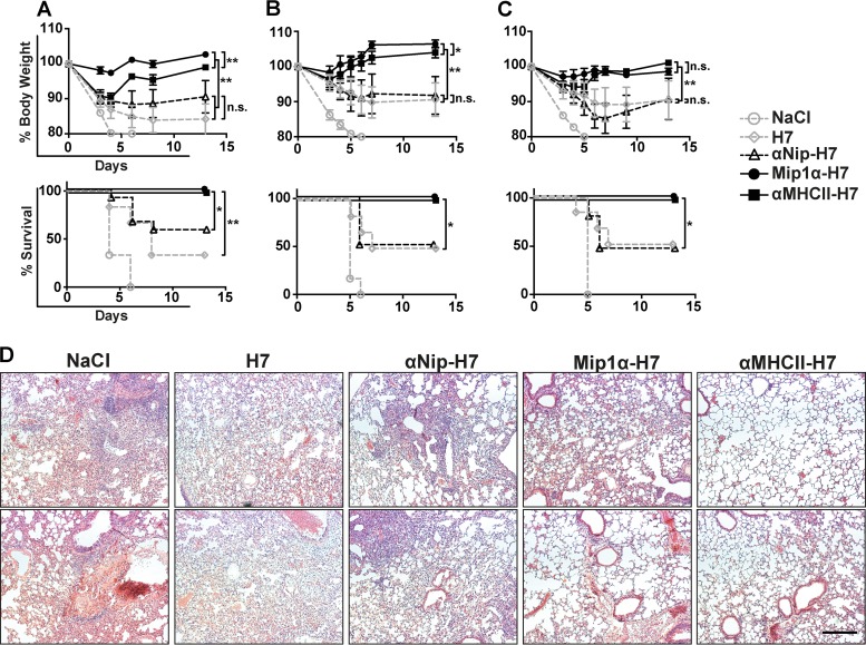
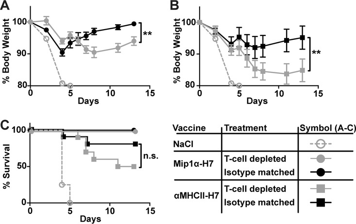
Zoonotic influenza H7 viral infections have a case fatality rate of about 40%. Currently, no or limited human to human spread has occurred, but we may be facing a severe pandemic threat if the virus acquires the ability to transmit between humans. Novel vaccines that can be rapidly produced for global distribution are urgently needed, and DNA vaccines may be the only type of vaccine that allows for the speed necessary to quench an emerging pandemic. Here, we constructed DNA vaccines encoding the hemagglutinin (HA) from influenza A/chicken/Italy/13474/99 (H7N1). In order to increase the efficacy of DNA vaccination, HA was targeted to either major histocompatibility complex class II molecules or chemokine receptors 1, 3, and 5 (CCR1/3/5) that are expressed on antigen-presenting cells (APC). A single DNA vaccination with APC-targeted HA significantly increased antibody levels in sera compared to nontargeted control vaccines. The antibodies were confirmed neutralizing in an H7 pseudotype-based neutralization assay. Furthermore, the APC-targeted vaccines increased the levels of antigen-specific cytotoxic T cells, and a single DNA vaccination could confer protection against a lethal challenge with influenza A/turkey/Italy/3889/1999 (H7N1) in mice. In conclusion, we have developed a vaccine that rapidly could contribute protection against a pandemic threat from avian influenza.IMPORTANCE Highly pathogenic avian influenza H7 constitute a pandemic threat that can cause severe illness and death in infected individuals. Vaccination is the main method of prophylaxis against influenza, but current vaccine strategies fall short in a pandemic situation due to a prolonged production time and insufficient production capabilities. In contrast, a DNA vaccine can be rapidly produced and deployed to prevent the potential escalation of a highly pathogenic influenza pandemic. We here demonstrate that a single DNA delivery of hemagglutinin from an H7 influenza could mediate full protection against a lethal challenge with H7N1 influenza in mice. Vaccine efficacy was contingent on targeting of the secreted vaccine protein to antigen-presenting cells.

Keywords: APC-targeting; DNA vaccine; avian viruses; hemagglutinin; influenza; pandemic influenza.


Figures

FIG 1
FIG 1
Characterization of vaccine proteins. (A) Schematic illustration of a dimeric vaccine protein. The targeting units, either an scFv specific for mouse I-Ed (αMHCII), the chemokine MIP1α (Mip1α), or a scFv specific for the hapten NIP (αNip; nontargeted control), are connected to HA via a dimerization unit containing the hinge and CH3 domain from human IgG3. (B) Western blot of supernatants from 293E cells transfected with the indicated vaccine plasmids. Molecular sizes and corresponding structures are indicated. (C) Vaccine proteins in supernatants from transiently transfected 293E cells were detected in an ELISA specific for HA from A/Shanghai/1/2013 (H7N9). (D) Binding of vaccine proteins to B cells (CD19+), macrophages (F4/80+ CD64+), DCs (Lin CD11chi) divided into conventional DC1 (CD24+) and conventional DC2 (CD11b+), and T cells (CD3+) from BALB/c splenocytes.
FIG 2
FIG 2
MHC-II-targeted HA vaccination with or without a multibasic cleavage site. (A) Alignment of the MBCS in H7 and the deleted corresponding sequence in H7Δ. (B) Schematic illustration depicting the vaccine monomer with indicated MBCS in HA. The full-length vaccine monomer is ∼150 kDa and HA2 is ∼35 kDa, resulting in fragments of ∼115 and ∼35 kDa, respectively, under reducing conditions. (C) Western blot of secreted vaccine proteins (indicated) under reducing conditions in supernatant from transiently transfected 293E cells. (D) Binding of secreted αMHCII-H7 or αMHCII-H7Δ proteins in supernatants from transiently transfected 293E cells detected in an ELISA specific for HA from A/Shanghai/1/2013 (H7N9). (E) Mice (n = 6/group) were vaccinated i.d. with plasmid DNA encoding either αMHCII-H7 or αMHCII-H7Δ, or NaCl, and IgG in sera measured in ELISA against recombinant HA from influenza A/Shanghai/1/2013(H7N9) at weeks 1, 2, and 4 postvaccination. *, P < 0.05 (two-tailed Mann-Whitney test).
FIG 3
FIG 3
Antibody responses after a single DNA vaccination. (A) Schematic illustration of the experiment. Briefly, BALB/c (n = 12/group) or CB6F1 mice (n = 6/group) were DNA vaccinated once i.d. with the indicated vaccine plasmids. Sera were collected up to 4 weeks postvaccination from BALB/c (B) and CB6F1 (C), and antibody responses were measured by ELISA against recombinant HA from influenza A/Shanghai/1/2013 (H7N9). *, P < 0.05; **, P < 0.01 (two-tailed Mann-Whitney test). (D) Sera from BALB/c from weeks 2 and 4 were pooled by group and assayed in a pseudotype microneutralization assay against A/FPV/Rostock/1934 (H7N1). Neutralization curves were fitted with GraphPad Prism 6 software, and the IC50 and IC90 titers were calculated. *, P < 0.05; **, P < 0.01 (extra sum-of-squares F test).
FIG 4
FIG 4
Induction of T cells after a single immunization. BALB/c mice (n = 6/group; n = 3/group for NaCl and H1 controls) were DNA vaccinated i.m. with plasmids encoding the indicated vaccines. Spleens were harvested at 8 weeks postvaccination. Splenocytes were restimulated with recombinant HA from A/Shanghai/1/2013(H7N9) (A) or HA from either A/Shanghai/1/2013(H7N9), A/Vietnam/1194/2004(H5N1), or A/Puerto Rico/8/34(H1N1) (B), and the numbers of IFN-γ-secreting cells were evaluated. (C) BALB/c mice (n = 6/group) were vaccinated i.d. with plasmid DNA encoding the indicated vaccines. After 5 weeks, 5 × 10⁶ A20 cells expressing cytosolic H7 and GFP and 5 × 10⁶ A20 cells expressing cytosolic mCherry were injected i.v. The prevalences of GFP- or mCherry-positive cells were assessed 16 h later, and the killing ratios were calculated. *, P < 0.05; **, P < 0.01 (two-tailed Mann-Whitney test).
FIG 5
FIG 5
Viral challenge of DNA-immunized mice. BALB/c mice (n = 6 to 12/group) or CB6F1 mice (n = 6/group) were vaccinated with 25 μg of DNA i.d. and challenged with 20 × LD50 of mouse-adapted A/turkey/Italy/3889/1999 (H7N1) at week 5 postvaccination. The body weight (upper panels) was measured after challenge to assess morbidity, and survival curves (lower panels) are shown for mice receiving the indicated vaccines from BALB/c mice (A) and CB6F1 mice (B). (C) BALB/c mice were vaccinated with vaccines encoding the HA antigen A/turkey/Italy/3889/1999 (H7N1) homologous to the challenge strain and challenged at week 5 postvaccination. Weight curves: *, P < 0.05; **, P < 0.01 (two-way ANOVA). Survival curves: *, P < 0.05; **, P < 0.01 (Gehan-Breslow-Wilcoxon test). (D) Representative micrographs of H&E-stained sections of lungs from each group from the experiment in panel A collected 7 days postchallenge. Scale bar, 250 μm.
FIG 6
FIG 6
Viral challenge of DNA-immunized mice after depletion of T cells. Mice (n = 8 to 10/group) were DNA vaccinated i.d. with the indicated vaccines and then challenged with 20 × LD50 A/turkey/Italy/3889/1999 (H7N1) 5 weeks postvaccination. Two days prior to challenge—and every other day until completion of the experiment—mice were injected i.p. with either CD4 and CD8 depleting MAbs or isotype-matched MAbs. Starting at the day of challenge (D0), weight was monitored in mice vaccinated with Mip1α-H7 (A) or αMHCII-H7 (B). *, P < 0.05; **, P < 0.01 (two-way ANOVA). (C) Survival curves for mice in panels A and B. *, P < 0.05; **, P < 0.01 (Gehan-Breslow-Wilcoxon test).

Similar articles

Cited by

References

    1. World Health Organization. 2017. Influenza at the human-animal interface, June 2017: monthly risk assessment summary, p 1–7 World Health Organization, Geneva, Switzerland.
    1. Fouchier RaM, Schneeberger PM, Rozendaal FW, Broekman JM, Kemink SaG, Munster V, Kuiken T, Rimmelzwaan GF, Schutten M, Van Doornum GJJ, Koch G, Bosman A, Koopmans M, Osterhaus ADME. 2004. Avian influenza A virus (H7N7) associated with human conjunctivitis and a fatal case of acute respiratory distress syndrome. Proc Natl Acad Sci U S A 101:1356–1361. doi:10.1073/pnas.0308352100. - DOI - PMC - PubMed
    1. Zanin M, Koçer ZA, Poulson RL, Gabbard JD, Howerth EW, Jones CA, Friedman K, Seiler J, Danner A, Kercher L, McBride R, Paulson JC, Wentworth DE, Krauss S, Tompkins SM, Stallknecht DE, Webster RG. 2016. The potential for low pathogenic avian H7 influenza a viruses to replicate and cause disease in a mammalian model. J Virol 91:e01934-16. doi:10.1128/JVI.01934-16. - DOI - PMC - PubMed
    1. Krammer F, Palese P. 2015. Advances in the development of influenza virus vaccines. Nat Rev Drug Discov 14:167–182. doi:10.1038/nrd4529. - DOI - PubMed
    1. Zhou L, Ren R, Yang L, Bao C, Wu J, Wang D, Li C, Xiang N, Wang Y, Li D, Sui H, Shu Y, Feng Z, Li Q, Ni D. 2017. Sudden increase in human infection with avian influenza A(H7N9) virus in China, September–December 2016. Western Pac Surveill Response J 8:6–14. doi:10.5365/wpsar.2017.8.1.001. - DOI - PMC - PubMed

MeSH terms