. 2017 Sep 21;2(18):e95352.
doi: 10.1172/jci.insight.95352.

Monocyte dysregulation and systemic inflammation during pediatric falciparum malaria

Affiliations

Monocyte dysregulation and systemic inflammation during pediatric falciparum malaria

Katherine R Dobbs et al. JCI Insight. .

Abstract

Background: Inflammation and monocytes are thought to be important to human malaria pathogenesis. However, the relationship of inflammation and various monocyte functions to acute malaria, recovery from acute malaria, and asymptomatic parasitemia in endemic populations is poorly understood.

Methods: We evaluated plasma cytokine levels, monocyte subsets, monocyte functional responses, and monocyte inflammatory transcriptional profiles of 1- to 10-year-old Kenyan children at the time of presentation with acute uncomplicated malaria and at recovery 6 weeks later; these results were compared with analogous data from asymptomatic children and adults in the same community.

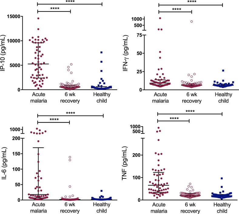
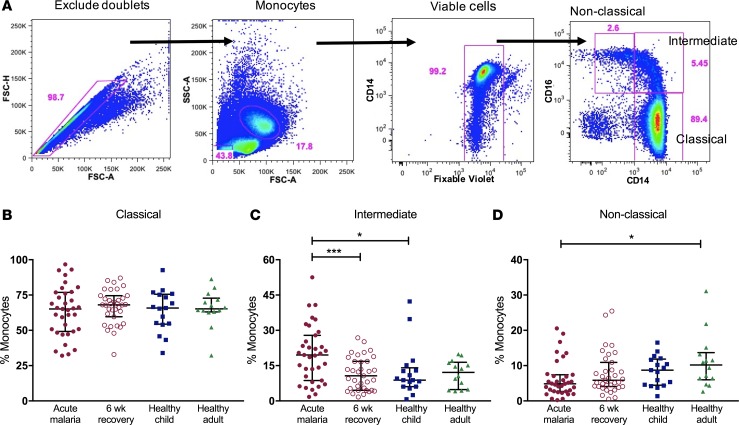
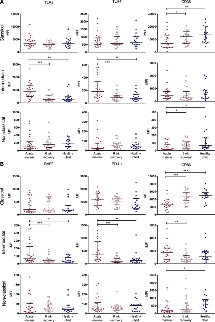
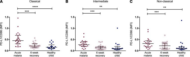
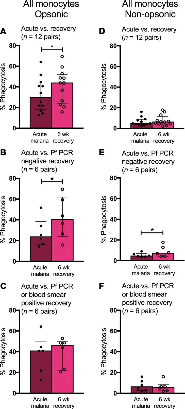
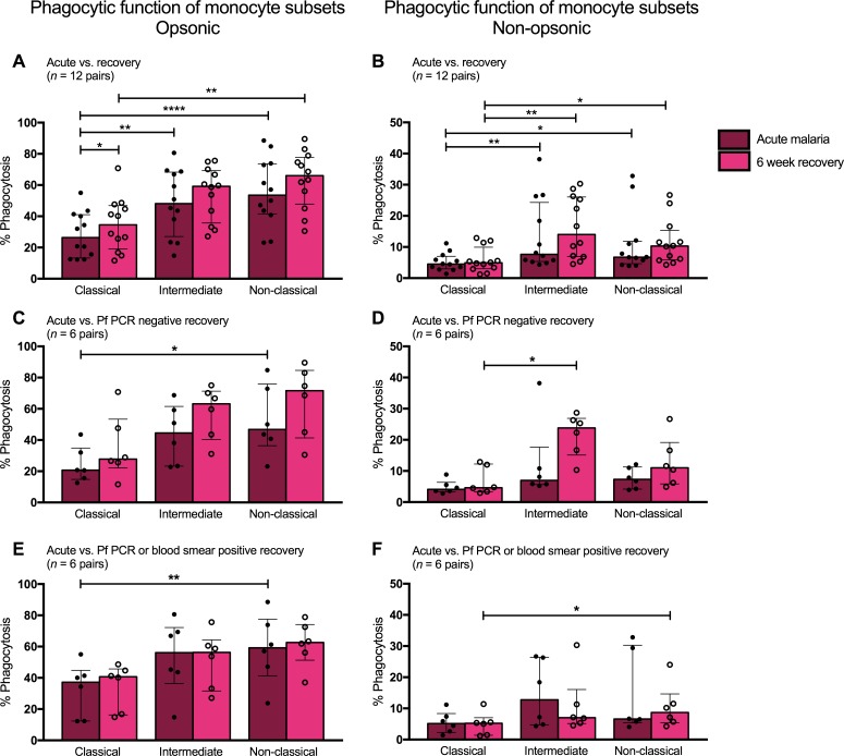
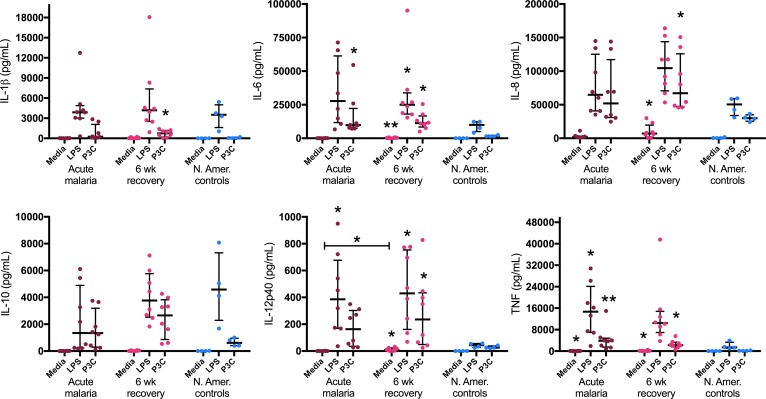
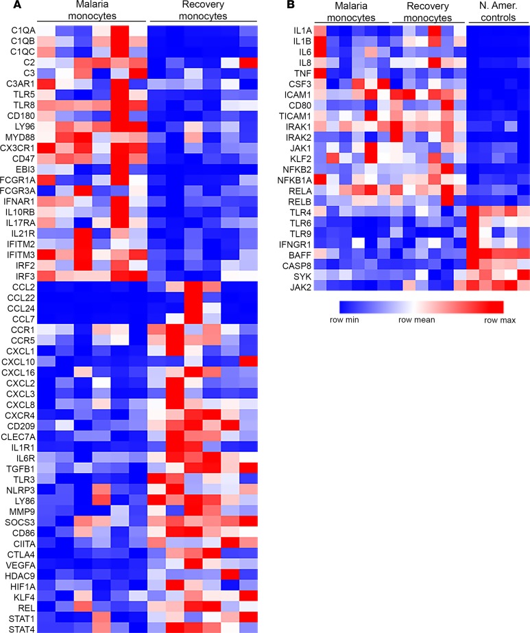
Results: Acute malaria was marked by elevated levels of proinflammatory and regulatory cytokines and expansion of the inflammatory "intermediate" monocyte subset that returned to levels of healthy asymptomatic children 6 weeks later. Monocytes displayed activated phenotypes during acute malaria, with changes in surface expression of markers important to innate and adaptive immunity. Functionally, acute malaria monocytes and monocytes from asymptomatic infected children had impaired phagocytosis of P. falciparum-infected erythrocytes relative to asymptomatic children with no blood-stage infection. Monocytes from both acute malaria and recovery time points displayed strong and equivalent cytokine responsiveness to innate immune agonists that were independent of infection status. Monocyte transcriptional profiles revealed regulated and balanced proinflammatory and antiinflammatory and altered phagocytosis gene expression patterns distinct from malaria-naive monocytes.

Conclusion: These observations provide insights into monocyte functions and the innate immune response during uncomplicated malaria and suggest that asymptomatic parasitemia in children is not clinically benign.

Funding: Support for this work was provided by NIH/National Institute of Allergy and Infectious Diseases (R01AI095192-05), the Burroughs Wellcome Fund/American Society of Tropical Medicine and Hygiene, and the Rainbow Babies & Children's Foundation.

Keywords: Immunology; Infectious disease; Innate immunity; Malaria; Monocytes.


Conflict of interest statement

Conflict of interest: The authors have declared that no conflict of interest exists.

Figures

Figure 1
Figure 1. Acute uncomplicated malaria is associated with a robust proinflammatory cytokine response and systemic inflammation.
Plasma cytokine and acute-phase reactant levels were compared in samples from cases of acute uncomplicated malaria and 6-week recovery samples (n = 60 pairs) and samples from heathy child controls (n = 40). Bars represent medians with interquartile ranges. Wilcoxon matched-pairs rank test was used to compare acute malaria samples to 6-week recovery samples. Kruskal-Wallis test was used to compare healthy child samples to acute malaria and 6-week recovery samples. ****P < 0.0001. IP-10, IFN-γ–induced protein 10.
Figure 2
Figure 2. Acute uncomplicated malaria is associated with an expansion of the intermediate monocyte subset.
(A) Gating strategy to calculate proportions of circulating monocyte subsets. Doublets were excluded, and monocytes were gated based on forward- and side-scatter properties. Dead cells were excluded based on Fixable Violet staining. The 3 subsets were determined based on CD14 and CD16 expression (classical CD14++CD16, intermediate CD14++CD16+, and nonclassical CD14+CD16++). Proportions of all circulating monocytes that are (B) classical, (C) intermediate, and (D) nonclassical for samples from cases of acute malaria, 6-week recovery (n = 35 pairs), healthy child controls (n = 17), and healthy adult controls (n = 14). Bars represent the median values with interquartile ranges. Wilcoxon matched-pairs rank test was used to compare acute malaria to 6-week recovery samples. Kruskal-Wallis test was used to compare unmatched groups. *P < 0.05, ***P < 0.001.
Figure 3
Figure 3. Cell surface phenotypes of monocyte subsets are activated during acute uncomplicated malaria.
Flow cytometry was used to determine cell surface expression of 6 markers on the three monocyte subsets (classical, intermediate, and nonclassical) for samples from cases of acute malaria, 6-week recovery samples (n = 23 pairs), and samples from healthy child controls (n = 17). (A) Cell surface expression of markers important in innate immunity: TLR2, TLR4, and CD36. (B) Cell surface expression of markers important in adaptive immunity: BAFF, PD-L1, and CD86. Wilcoxon matched-pairs rank test was used to compare acute malaria samples to 6-week recovery samples. Kruskal-Wallis test was used to compare healthy child samples to acute malaria and 6-week recovery samples. Bars represent the median values with interquartile ranges. *P < 0.05, **P < 0.01, ***P < 0.001, ****P < 0.0001. iMFI, integrated mean fluorescence intensity; BAFF, B cell–activating factor; PD-L1, programmed death-ligand 1.
Figure 4
Figure 4. Acute uncomplicated malaria is associated with an increased PD-L1/CD86 ratio on all monocyte subsets.
Ratios of cell surface expression of PD-L1/CD86 on the (A) classical, (B) intermediate, and (C) nonclassical monocyte subsets for samples from cases of acute malaria, 6-week recovery samples (n = 23 pairs), and samples from healthy child controls (n = 17) are indicated on the y axes. Bars represent the median values with interquartile ranges. Wilcoxon matched-pairs rank test was used to compare acute malaria samples to 6-week recovery samples. Kruskal-Wallis test was used to compare healthy children to acute malaria samples and 6-week recovery samples. **P < 0.01, ***P < 0.001, ****P < 0.0001. iMFI, integrated mean fluorescence intensity; PD-L1, programmed death-ligand 1.
Figure 5
Figure 5. Monocytes display decreased phagocytic function during P. falciparum infection.
(A) Opsonic phagocytic function of all monocytes from acute malaria (AM) and 6-week recovery PBMC samples (n = 12 pairs) in the presence of Ghana14 IEs opsonized with heat-inactivated pooled plasma from Kenyan adults. (B) Opsonic phagocytic function of all monocytes from AM and 6-week PBMC samples (n = 6 pairs) from a subset of children who were P. falciparum PCR negative at 6-week follow up. (C) Opsonic phagocytic function of all monocytes from AM and 6-week PBMC samples (n = 6 pairs) from a subset of children with asymptomatic P. falciparum infection at 6-week follow up (defined by positive blood smear or P. falciparum PCR). (D) Nonopsonic phagocytic function of all monocytes from AM and 6-week PBMC samples (n = 12 pairs) in the presence of Ghana14 IEs opsonized with heat-inactivated plasma from a malaria-naive North American. (E) Nonopsonic phagocytic function of all monocytes from AM and 6-week PBMC samples (n = 6 pairs) from a subset of children who were P. falciparum PCR negative at 6-week follow up. (F) Nonopsonic phagocytic function of all monocytes from AM and 6-week PBMC samples (n = 6 pairs) from a subset of children with asymptomatic P. falciparum infection at 6-week follow up (defined by positive blood smear or P. falciparum PCR). Wilcoxon matched-pairs rank test was used to compare AM to 6-week recovery samples. Data are shown as medians with interquartile ranges. *P < 0.05.
Figure 6
Figure 6. Intermediate and nonclassical monocytes display greater phagocytic function compared with classical monocytes.
(A) Opsonic phagocytic function of the three monocyte subsets from acute malaria (AM) and 6-week recovery PBMC samples (n = 12 pairs) in the presence of Ghana14 IEs opsonized with heat-inactivated pooled plasma from malaria-immune Kenyan adults. (B) Nonopsonic phagocytic function of monocyte subsets from AM and 6-week PBMC samples (n = 12 pairs) in the presence of Ghana14 IEs opsonized with heat-inactivated plasma from a malaria-naive North American. (C) Opsonic phagocytic function of monocyte subsets from AM and 6-week PBMC samples (n = 6 pairs) from a subset of children who were P. falciparum PCR negative at 6 weeks. (D) Nonopsonic phagocytic function of monocyte subsets from AM and 6-week PBMC samples (n = 6 pairs) from a subset of children who were P. falciparum PCR negative at 6 weeks. (E) Opsonic phagocytic function of monocyte subsets from AM and 6-week PBMC samples (n = 6 pairs) from a subset of children with asymptomatic P. falciparum infection at 6 weeks (defined by positive blood smear or P. falciparum PCR). (F) Nonopsonic phagocytic function of monocyte subsets from AM and 6-week PBMC samples (n = 6 pairs) from a subset of children with asymptomatic P. falciparum infection at 6 weeks (defined by positive blood smear or P. falciparum PCR). Differences between AM and 6-week PBMC samples analyzed by Wilcoxon matched-pairs rank test. Differences among subsets analyzed by Friedman test with multiple comparisons. Data shown are medians with interquartile ranges. *P < 0.05, **P < 0.01, ****P < 0.0001.
Figure 7
Figure 7. Monocytes from children during acute malaria and 6 weeks following treatment are highly responsive to stimulation with TLR ligands.
Monocytes were negatively selected from fresh venous blood samples from children during acute malaria and 6 weeks following treatment (n = 8 pairs). Healthy North American (N. Amer.) adult controls (n = 4) were used as experimental controls. All assays were performed with technical duplicates. Cells were cultured for 18 hours with media alone, 10 ng/ml LPS, or 100 ng/ml Pam3CSK4 (P3C), and cytokine concentrations were measured in culture supernatants. Wilcoxon matched-pairs rank test was used to compare acute malaria to 6-week recovery samples. Kruskal-Wallis test was used to compare acute malaria, 6-week recovery, and N. Amer. control samples. Data are shown as medians with interquartile ranges. *P < 0.05, **P < 0.01, as compared with controls.
Figure 8
Figure 8. Monocytes from children with uncomplicated malaria have a distinct, regulated inflammatory gene expression profile.
Targeted digital RNA sequencing was performed on monocytes isolated from cryopreserved PBMCs via negative selection over a magnetic column. A customized panel targeted 508 genes important in inflammation and immunity (QIAseq Human Inflammation and Immunity Transcriptome Panel, Qiagen). Differential expression was determined using the DESeq2 R/Bioconductor package. (A) Monocyte transcriptional profiles were compared between samples from cases of acute malaria and matching 6-week recovery samples (n = 6 pairs). Of the 508 genes analyzed, 125 were differentially expressed between AM and 6-week recovery (51 overexpressed and 74 underexpressed during AM; P < 0.05). Key genes are listed in the heatmap. (B) Monocyte transcriptional profiles were compared among samples from children with acute malaria, 6-week recovery samples (n = 6 pairs), and samples from healthy malaria-naive North American controls (n = 5). Of the 508 genes analyzed, 114 were overexpressed and 86 underexpressed during acute malaria compared with North American controls and 124 were overexpressed and 95 underexpressed at 6-week recovery compared with North American (N. Amer.) controls (P < 0.05). Key genes are listed in the heatmap. None of the genes listed in B were differentially expressed between acute malaria and recovery.

Similar articles

Cited by

References

    1. World Malaria Report 2015. World Health Organization. http://www.who.int/malaria/publications/world-malaria-report-2015/report... Accessed August 24, 2017.
    1. Imbert P, et al. Severe falciparum malaria in children: a comparative study of 1990 and 2000 WHO criteria for clinical presentation, prognosis and intensive care in Dakar, Senegal. Trans R Soc Trop Med Hyg. 2002;96(3):278–281. doi: 10.1016/S0035-9203(02)90099-4. - DOI - PubMed
    1. Reyburn H, et al. Association of transmission intensity and age with clinical manifestations and case fatality of severe Plasmodium falciparum malaria. JAMA. 2005;293(12):1461–1470. doi: 10.1001/jama.293.12.1461. - DOI - PubMed
    1. Kendjo E, et al. Mortality patterns and site heterogeneity of severe malaria in African children. PLoS One. 2013;8(3):e58686. doi: 10.1371/journal.pone.0058686. - DOI - PMC - PubMed
    1. Langhorne J, Ndungu FM, Sponaas AM, Marsh K. Immunity to malaria: more questions than answers. Nat Immunol. 2008;9(7):725–732. doi: 10.1038/ni.f.205. - DOI - PubMed

Publication types