. 2017 Sep 21;2(18):e95530.
doi: 10.1172/jci.insight.95530.

Reversible retinal vessel closure from VEGF-induced leukocyte plugging

Affiliations

Reversible retinal vessel closure from VEGF-induced leukocyte plugging

Yuanyuan Liu et al. JCI Insight. .

Abstract

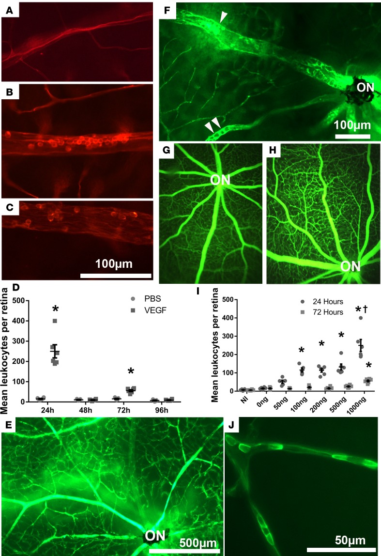
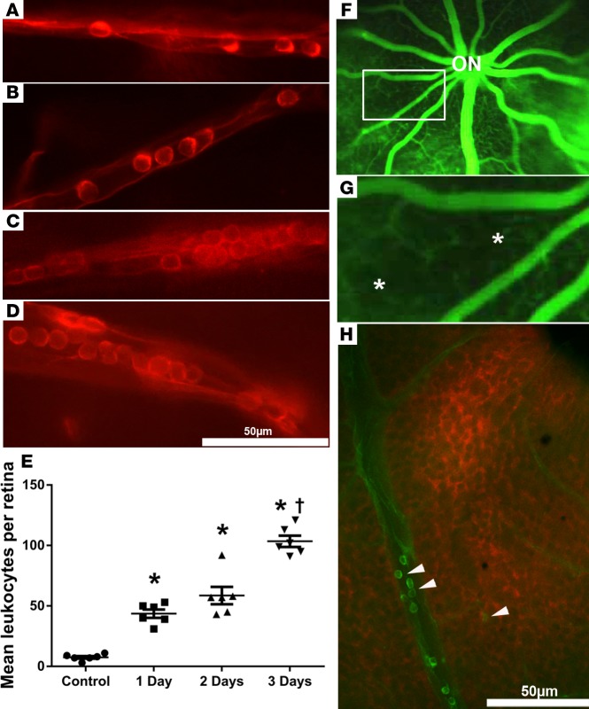
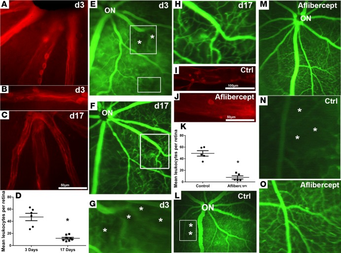
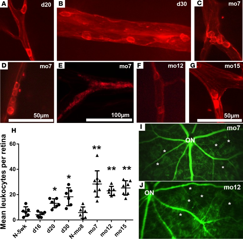
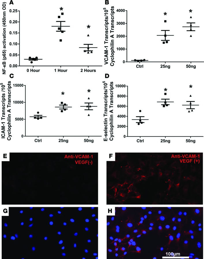
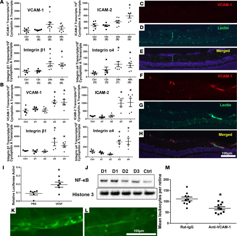
Clinical trials in patients with macular edema due to diabetic retinopathy or retinal vein occlusion (RVO) have shown that suppression of VEGF not only improves macular edema, but also reopens closed retinal vessels, prevents progression of vessel closure, and improves retinopathy. In this study, we show the molecular basis for those clinical observations. Increased retinal levels of VEGF in mice cause plugging of retinal vessels with leukocytes, vessel closure, and hypoxia. Suppression of VEGF reduces leukocyte plugging, causing reperfusion of closed vessels. Activation of VEGFR1 contributes to leukocyte recruitment, because it is significantly reduced by an anti-VEGFR1-neutralizing antibody. High VEGF increases transcriptional activity of NF-κB and expression of NF-κB target genes, particularly Vcam1. Injection of an anti-VCAM-1-neutralizing antibody reduces VEGF-induced leukocyte plugging. These data explain the broad range of benefits obtained by VEGF suppression in patients with ischemic retinopathies, provide an important insight into the pathogenesis of RVO and diabetic retinopathy, and suggest that sustained suppression of VEGF early in the course of these diseases may prevent vessel closure, worsening ischemia, and disease progression. This study also identifies VEGFR1 and VCAM-1 as molecular targets whose suppression could supplement VEGF neutralization for treatment of RVO and diabetic retinopathy.

Keywords: Retinopathy; Vascular Biology; endothelial cells; hypoxia.


Conflict of interest statement

Conflict of interest: The authors have declared that no conflict of interest exists.

Figures

Figure 1
Figure 1. Intravitreous injection of VEGF causes transient severe leukostasis in retinal vessels.
Retinal vessels in mice perfused with rhodamine-labeled concanavalin A (con A) were narrow and most did not contain any leukocytes 24 hours after intravitreous injection of PBS (A) but were dilated and packed with leukocytes 24 hours (B) or 72 hours (C) after intravitreous injection of 1 μg VEGF. The mean (± SEM) number of intravascular leukocytes per retina (PBS-treated mice, n = 5; VEGF-treated mice, n = 6 at each time point) was significantly greater in VEGF-injected eyes compared with PBS-injected eyes at 24 and 72 hours after injection (*P < 0.001 by unpaired t tests) but not at 48 or 96 hours (D, P = 0.6179 [48 hours], P = 0.4722 [96 hours] by unpaired t tests). Retinal vessels in a region around the optic nerve (ON) of mice perfused with fluorescein-labeled dextran showed normal retinal vasculature 24 hours after intravitreous injection of PBS (E), whereas vessels were dilated and packed with leukocytes seen in negative relief 24 hours after injection of 1 μg VEGF (F, arrowheads). Fluorescein angiography 24 hours after injection of PBS (G) or 1 μg VEGF (H) showed no identifiable nonperfusion. (I) The mean (± SEM) number of intravascular leukocytes per retina (NI group n = 6, mice treated with 200 ng, 500 ng, 1,000 ng VEGF n = 6; mice treated with 0 ng, 50 ng, 100 ng VEGF n = 5) was determined for several doses of VEGF and was significantly greater than PBS control for doses ≥100 ng (*P ≤ 0.002, P < 0.001; P = 0.002 [100 ng, 24 hours], P = 0.001 [200 ng, 24 hours], P < 0.001 [500 ng, 24 hours], P < 0.001 [1,000 ng, 24 hours and 72 hours] by 1-way ANOVA with Bonferroni correction for multiple comparisons). (J) Twenty-four hours after intravitreous injection of 200 ng VEGF, perfusion with fluorescein-labeled Con A showed relatively small retinal vessels plugged with leukocytes. Scale bar: 50 μm (J); 100 μm (AC and F); 500 μm (E).
Figure 2
Figure 2. Sustained expression of VEGF in the retina causes leukostasis, retinal vessel closure, and retinal hypoxia.
Tet/opsin/VEGF double-transgenic mice with doxycycline-inducible expression of VEGF in the retina were given 2 mg/ml doxycycline in drinking water and perfused with rhodamine-labeled Con A 1 day (A), 2 days (B), or 3 days (C and D) after initiating doxycycline. Leukocytes were present in small vessels 1 day (A) and 2 days (B) after onset of VEGF expression and were present in vessels of all sizes after 3 days (C and D). The mean (± SEM) number of intravascular leukocytes per retina was significantly greater at each of the 3 time points compared with control (E) (*P < 0.001 by 1-way ANOVA with Bonferroni corrections) and at day 3 was greater than other time points (P < 0.01, 1-way ANOVA with Bonferroni corrections). (F) Fluorescein angiography of Tet/opsin/VEGF mice 3 days after starting doxycycline showed dilated large retinal vessels radiating from the optic nerve (ON), between which the networks of small vessels were slightly blurred by extravascular leakage. There were hypofluorescent areas with sharp borders that appeared cut out of the diffuse fluorescence (box), better seen in a magnified view of the boxed area (G, asterisks). The dark black areas indicate absence of capillaries due to nonperfusion. Staining of a retinal flat mount with hydroxyprobe 3 days after starting doxycycline showed hypoxic retina (red) adjacent to vessels containing adherent FITC–Con A–stained leukocytes (H, arrowheads). Scale bar: 50 μm.
Figure 3
Figure 3. Turning off VEGF expression or injection of a VEGF-neutralizing protein reduces leukostasis and opens closed vessels.
On day 3 after intraperitoneal injections of doxycycline on day 0 (50 mg/kg), day 1 (25 mg/kg), and day 2 (25 mg/kg), retinal flat mounts from rhodamine-labeled concanavalin A–perfused (Con A–perfused) Tet/opsin/VEGF mice showed leukocytes adherent to the walls of retinal vessels (A) that appeared to plug the lumen of some vessels (B). Other Tet/opsin/VEGF mice were given 3 injections of doxycycline and then maintained without treatment until day 17 when they were perfused with rhodamine-labeled Con A; retinal flat mounts showed few intravascular leukocytes (C). Tet/opsin/VEGF mice treated in the same manner were perfused with rhodamine-labeled Con A on day 3 or day 17 (n = 6, n=8), and the mean (± SEM) number of intravascular leukocytes per retina was lower on day 17 than on day 3 (D, *P < 0.001 by unpaired t test), indicating that most of the leukocytes had cleared. (E). Fluorescein angiograms obtained on day 3 showed dilation of large retinal vessels between which there was continuous fluorescence interrupted by hypofluorescent regions of nonperfusion (boxes, asterisks). (F). A repeat fluorescein angiogram in the same Tet/opsin/VEGF mouse on day 17 showed that the large retinal vessels were no longer dilated, leakage had resolved allowing better resolution of small vessels, and the previously black areas were filled in with a continuous network of vessels, indicating reopening of previously closed vessels. Magnified images of the large boxed areas in E and F allowed a better visualization of regions of vessel closure induced by high expression of VEGF (G, asterisks), while the same region of retina showed reperfused vessels after levels of VEGF returned to normal (H).Tet/opsin/VEGF mice were given an intravitreous injection of 40 μg aflibercept (n = 5) or PBS (n = 5), and 2 mg/ml doxycycline was added to their drinking water. After 3 days, control mice showed numerous intravascular leukocytes (I), while few leukocytes were seen in vessels of aflibercept-injected eyes (J). The mean (± SEM) number of intravascular leukocytes was significantly lower in eyes injected with aflibercept (K, *P < 0.001 for difference from control by unpaired t test). Fluorescein angiograms in control eyes showed dilation of large retinal vessels, diffuse hyperfluorescence from extravascular leakage, and areas of retinal nonperfusion (L, box), whereas eyes injected with aflibercept showed no vessel dilation, leakage, or retinal nonperfusion (M). Magnification of the boxed area in L clearly shows the dark black patches, indicating closure of retinal vessels in control mice (N, Ctrl, asterisks), while the magnified view of the retinal vessels in M shows the absence of any vessel closure in aflibercept-treated mice (O).
Figure 4
Figure 4. Chronic elevation of VEGF in the retinas of rho/VEGF-transgenic mice causes gradually increasing leukostasis and retinal nonperfusion.
Rho/VEGF-transgenic mice of different ages, day 16, day 20, day 30, 7 months, 12 months, or 15 months, or normal wild-type mice at 5 weeks (N-5wk) or 8 months of age (N-mo8) were perfused with rhodamine-labeled Con A. Several vessels showed single adherent leukocytes in day 20 rho/VEGF mice (A) and more were seen in day 30 mice (B). In 7-month-old or older rho/VEGF mice, there were many more intravascular leukocytes and they often appeared to occlude the lumen of vessels exemplified in 3 separate 7-month-old mice by plugging at a vessel bifurcation (C), by a line of leukocytes (D), or packing of a substantial length of a vessel lumen with a large aggregate (E). Similarly, there were leukocytes plugging vessels particularly at branch points in the retinas of 12-month-old (F) and 15-month-old (G) rho/VEGF mice. The mean (± SEM) number of intravascular leukocytes per retina in both 20-day-old and 30-day-old rho/VEGF mice, but not 16-day-old mice, was significantly greater than that in N-5W control mice (*P = 0.05; P = 0.022 [day 20], P < 0.001 [day 30], P = 0.349 [day 16] compared to 5 weeks by unpaired t tests) and mean intravascular leukocytes were significantly greater in 7-month-old, 12-month-old, and 15-month-old rho/VEGF mice compared with N-8M control mice (H) (n = 6 for each bar except 7 months, for which n = 9; **P < 0.001 by unpaired t tests). Retinal flat mounts from fluorescein-dextran-perfused 7-month-old (I) and 12-month-old (J) rho/VEGF mice show abruptly ending vessels and large areas of nonperfusion (asterisks). Scale bar: 50 μm (AD and G); 100 μm (E and F).
Figure 5
Figure 5. Intravitreous injection of a VEGF-neutralizing protein causes opening of some vessels closed by chronic overexpression of VEGF in the retinas of rho/VEGF mice.
Fluorescein angiography (FA) was done on 7-month-old rho/VEGF mice (A), and then the mice were given subcutaneous injections of 25 mg/kg aflibercept every other day for a total of 4 injections followed by repeat FA (B). At baseline prior to aflibercept injection, several black areas devoid of fluorescein were seen, indicating closure of retinal vessels (A, asterisks and dots within boxes). After aflibercept injections, many of the nonperfused areas were perfused indicating opening of previously closed vessels (B, boxes). Comparison of a magnified view of the large white box in A (C) with a magnified view of box in B, the same region of the retina after aflibercept treatment (D), shows 4 black regions devoid of blood vessels (C, asterisks) in which vessels reappear after aflibercept treatment indicating reopening (D). However, other black areas (dots) remain black after aflibercept treatment indicating that not all closed vessels reopen.
Figure 6
Figure 6. VEGFR1 contributes to leukocyte recruitment.
C57BL/6 mice were given a tail vein injection of 100 μg rat anti-mouse VEGFR1 or rat IgG and then an intravitreous injection of 200 ng VEGF; 24 hours later, mice were perfused with FITC-labeled Con A. There were many intravascular leukocytes in rat IgG-injected mice (A) and significantly fewer in those injected with anti-VEGFR1 (B and C) (n = 6 for rat IgG and n = 10 for anti-VEGFR1; *P = 0.0069 for difference from rat IgG by unpaired t test). Twenty-four hours after intravitreous injection of 200 ng VEGF, C57BL/6 mice were perfused with FITC-labeled Con A (green) and retinal flat mounts were counterstained (red) with anti-F4/80 (D–F) or anti-Ly6G (G–I). Scale bar: 50 μm (DI).
Figure 7
Figure 7. VEGF stimulates transcriptional activity of NF-κB and increases expression of adhesion molecules in retinal vascular endothelial cells.
(A) Primary human retinal endothelial cells (HRECs) were incubated in 25 ng/ml VEGF for 0, 1, or 2 hours (n = 5 wells for each time point), and 3.5 μg nuclear protein extract from each well was used to measure NF-κB transcriptional activity, as described in Methods. Lines represent the mean (± SEM) optical density (OD) at 450 nm. *P < 0.001, P = 0.032 [2 hours] for comparison with unincubated controls by unpaired t tests. (B–D) HRECs were incubated in 0, 25, or 50 ng/ml VEGF for 4 hours (n = 4 triplicate wells per group), and then mRNA for adhesion molecules was measured by qRT-PCR and normalized to mRNA for Cyclophilin A for normalization. Lines represent mean (± SEM) mRNA copies of target gene per 105 copies of Cyclophilin A mRNA. *P < 0.05 for comparison with unincubated controls by unpaired t tests. (E–H) Primary mouse retinal endothelial cells were incubated in the absence (VEGF-) or presence of 25 ng/ml VEGF for 4 hours and then immunostained with rat anti-mouse VCAM-1 and goat anti-rat secondary antibody conjugated with Alexa Fluor 594 (red) and counterstained with Hoechst (blue); there was more VCAM-1 staining in VEGF-treated cells. Scale bar: 100 μm.
Figure 8
Figure 8. VEGF stimulates expression of VCAM-1 in the retina, and neutralization of VCAM-1 reduces VEGF-induced leukostasis.
(A) C57BL/6 mice were given an intravitreous injection of PBS or 1 μg VEGF, and qRT-PCR was used to measure retinal mRNA levels for several adhesion molecules. Compared with PBS controls, there was significant elevation of the mean (± SEM) number of mRNA transcripts per 105 Cyclophilin A transcripts for VCAM-1, ICAM-2, integrin β1, and integrin α4 (n in parentheses along x axis, *P < 0.05 by unpaired t tests). (B) In Tet/opsin/VEGF mice, the mean (± SEM) number of mRNA transcripts per 105 Cyclophilin A transcripts for VCAM-1, ICAM-2, integrin β1, and integrin α4 first became significantly elevated after 3 days of doxycycline treatment compared with controls not treated with doxycycline (n = 5 *P < 0.05 by unpaired t tests ). (C–H) One day after C57BL/6 mice were given an intravitreous injection of 1 μg VEGF in one eye and PBS in the fellow eye, ocular frozen sections immunostained for VCAM-1 (red) and Griffonia simplicifolia lectin (green) showed no detectable staining for VCAM-1 in PBS-injected eyes (C–E), but eyes that had been injected with VEGF showed several vessels that stained for VCAM-1 (F–H). (I) Fourteen days after subretinal injection of 0.5 μg NF-κB luciferase reporter vector and 0.25 ng Renilla luciferase plasmid in C57BL/6 mice, an intravitreous injection of 1 μg VEGF was performed. Five days later, the ratio of Firefly/Renilla luciferase was measured in retinal/choroidal homogenates by Dual-Luciferase Reporter Assay. Lines show that the mean relative luciferase activity (n = 6) was significantly greater in VEGF-injected eyes compared with PBS-injected eyes (*P = 0.0138 by unpaired t test). (J) Immunoblots of nuclear fractions from Tet/opsin/VEGF mice treated with doxycycline for 1 (D1), 2 (D2), or 3 days (D3) showed higher levels of NF-κB but equivalent histone 3 in nuclei from retinas with high levels of VEGF compared with those in untreated Tet/opsin/VEGF mice with low levels of VEGF (Ctrl). (K–M) Tet/opsin/VEGF mice (n = 10 for each group) were given an intravenous injection of 100 μg anti-mouse VCAM-1 antibody or rat IgG; after 3 days of doxycycline treatment, there were many adherent leukocytes in retinal vessels of IgG-injected mice (K) and significantly fewer in eyes of anti-VCAM-1–injected mice (L and M) (*P = 0.0013 by unpaired t test). Scale bar: 100 μm.

Similar articles

Cited by

References

    1. Yau JW, et al. Global prevalence and major risk factors of diabetic retinopathy. Diabetes Care. 2012;35(3):556–564. doi: 10.2337/dc11-1909. - DOI - PMC - PubMed
    1. Diabetes Control Complications Trial Research Group. et al. The effect of intensive treatment of diabetes on the development and progression of long-term complications in insulin-dependent diabetes mellitus. N Engl J Med. 1993;329(14):977–986. doi: 10.1056/NEJM199309303291401. - DOI - PubMed
    1. [No authors listed] Intensive blood-glucose control with sulphonylureas or insulin compared with conventional treatment and risk of complications in patients with type 2 diabetes (UKPDS 33). UK Prospective Diabetes Study (UKPDS) Group. Lancet. 1998;352(9131):837–853. doi: 10.1016/S0140-6736(98)07019-6. - DOI - PubMed
    1. Klein R, Knudtson MD, Lee KE, Gangnon R, Klein BE. The Wisconsin Epidemiologic Study of Diabetic Retinopathy XXIII: the twenty-five-year incidence of macular edema in persons with type 1 diabetes. Ophthalmology. 2009;116(3):497–503. doi: 10.1016/j.ophtha.2008.10.016. - DOI - PMC - PubMed
    1. Stitt AW. The role of advanced glycation in the pathogenesis of diabetic retinopathy. Exp Mol Pathol. 2003;75(1):95–108. doi: 10.1016/S0014-4800(03)00035-2. - DOI - PubMed

Publication types

MeSH terms

Substances