. 2017 Sep 21;2(18):e95918.
doi: 10.1172/jci.insight.95918.

Functional correction of dystrophin actin binding domain mutations by genome editing

Affiliations

Functional correction of dystrophin actin binding domain mutations by genome editing

Viktoriia Kyrychenko et al. JCI Insight. .

Abstract

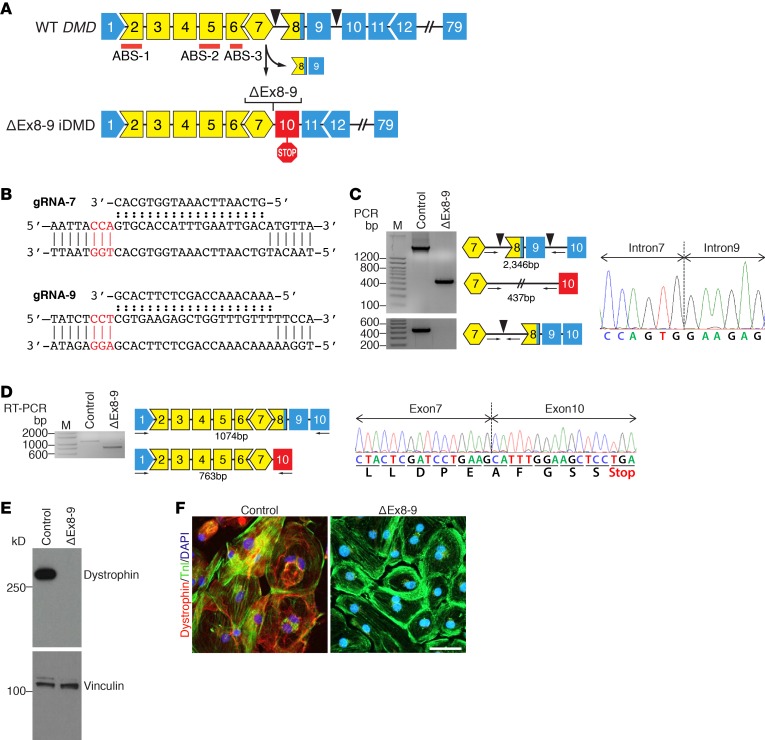
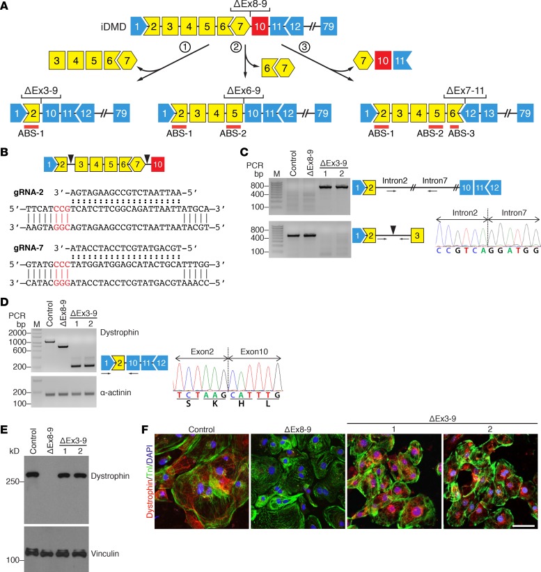
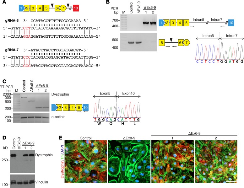
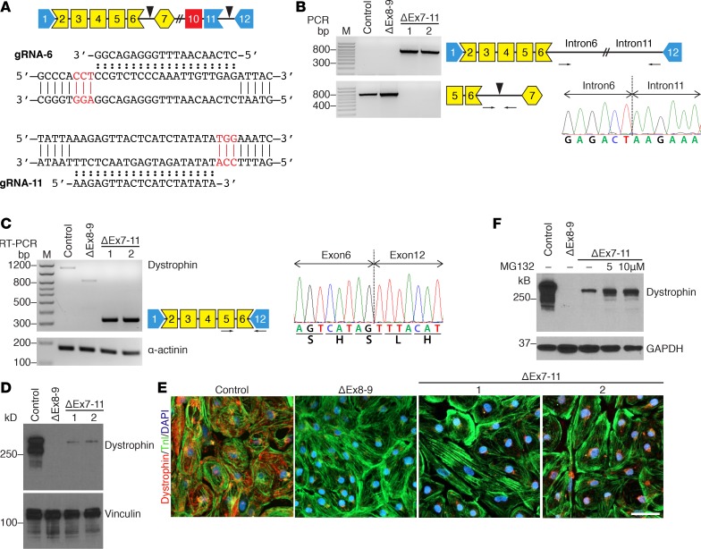
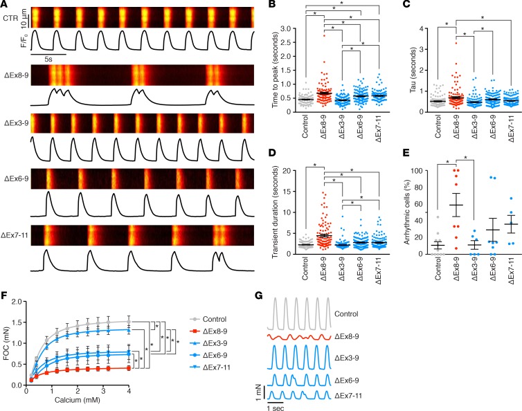
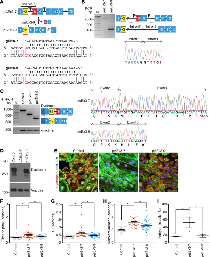
Dystrophin maintains the integrity of striated muscles by linking the actin cytoskeleton with the cell membrane. Duchenne muscular dystrophy (DMD) is caused by mutations in the dystrophin gene (DMD) that result in progressive, debilitating muscle weakness, cardiomyopathy, and a shortened lifespan. Mutations of dystrophin that disrupt the amino-terminal actin-binding domain 1 (ABD-1), encoded by exons 2-8, represent the second-most common cause of DMD. In the present study, we compared three different strategies for CRISPR/Cas9 genome editing to correct mutations in the ABD-1 region of the DMD gene by deleting exons 3-9, 6-9, or 7-11 in human induced pluripotent stem cells (iPSCs) and by assessing the function of iPSC-derived cardiomyocytes. All three exon deletion strategies enabled the expression of truncated dystrophin protein and restoration of cardiomyocyte contractility and calcium transients to varying degrees. We show that deletion of exons 3-9 by genomic editing provides an especially effective means of correcting disease-causing ABD-1 mutations. These findings represent an important step toward eventual correction of common DMD mutations and provide a means of rapidly assessing the expression and function of internally truncated forms of dystrophin-lacking portions of ABD-1.

Keywords: Gene therapy; Genetic diseases; Muscle; Muscle Biology.


Conflict of interest statement

Conflict of interest: RBD and ENO are consultants for Exonics Therapeutics. WHZ is a cofounder and, together with MT, scientific advisor of Myriamed.

Figures

Figure 1
Figure 1. Generating an iDMD model by deleting DMD exons 8–9 using CRISPR/Cas9-mediated genome editing.
(A) Strategy showing CRISPR/Cas9-mediated genomic editing of WT DMD to generate ΔEx8-9 iDMD. Shape and color of boxes denoting DMD exons indicate reading frame and protein coding domains. Yellow designates actin binding domain-1 (ABD-1). Blue marks part of the central rod domain. Red lines indicate actin binding sites (ABS1, ABS2, and ABS3). Arrowheads mark targeting site of guide RNAs (gRNAs). Stop sign marks exon with stop codon. (B) Sequences of gRNAs and their targeting sites within intron 7 (top) and intron 9 (bottom). gRNAs were designed to target the 3′ region of intron 7 (gRNA-7) and 5′ region of intron 9 (gRNA-9). PAM sites are highlighted in red. (C) PCR genotyping of control and ΔEx8-9 iDMD induced pluripotent stem cell (iPSC) lines using primers upstream and downstream of the gRNA targeting sites (top) and within intron 7 flanking the gRNA-7 targeting site (bottom). Sequencing of PCR product of ΔEx8-9 iDMD validates splicing of intron 7 to intron 9. PCR primers are indicated by arrows. Arrowhead indicates gRNA targeting site. M denotes marker lane. (D) RT-PCR analysis of dystrophin mRNA expression in control and ΔEx8-9 iDMD iPSC–derived cardiomyocytes. Forward primer targeting exon 1 and reverse primer targeting exon 10 were used. Sequencing confirmed splicing of exon 7 to exon 10, introducing a stop-codon. (E) Western blot analysis showing dystrophin protein expression in iPSC-derived cardiomyocytes using anti-dystrophin antibody. Vinculin was used as loading control. n = 7 for each group. (F) Immunocytochemistry representations of iPSC-derived cardiomyocytes with anti-dystrophin (red) and anti–troponin I (green). Nuclei are stained with Hoechst 33342 (blue). Scale bar: 50 μm. n = 4 for each group.
Figure 2
Figure 2. Correcting ΔEx8-9 iDMD by exon deletion to restore dystrophin expression.
(A) Three strategies were used to correct ΔEx8-9 iDMD by CRISPR/Cas9-mediated genomic editing: (i) deleting exons 3–7 to generate ΔEx3-9, (ii) deleting exons 6–7 to generate ΔEx6-9, and (iii) deleting exons 7–11 to generate ΔEx7-11. Shape and color of boxes denoting DMD exons indicate reading frame and protein coding domains. Yellow designates actin binding domain-1 (ABD-1). Blue marks part of the central rod domain. Red lines indicate actin binding sites (ABS1, ABS2, and ABS3). Red exon indicates exon with stop codon. (B) Illustration showing deletion of exons 3–7 to generate ΔEx3-9. Sequences of gRNAs and their targeting sites within intron 2 (top) and intron 7 (bottom). gRNAs were designed to target 3′ region of intron 2 (gRNA-2) and 5′ region of intron 7 (gRNA-7). Arrowheads mark targeting site of gRNAs. PAM sites are highlighted in red. (C) PCR genotyping of control, ΔEx8-9 iDMD, and two clones of ΔEx3-9 induced pluripotent cell (iPSC) lines using primers upstream and downstream of the gRNA targeting sites (top) and within intron 2 flanking the gRNA-2 targeting site (bottom). Sequencing of PCR product of ΔEx3-9 validates splicing of intron 2 to intron 7. PCR primers are indicated by arrows. Arrowhead indicates gRNA targeting site. M denotes marker lane. (D) RT-PCR analysis of dystrophin mRNA expression in control, ΔEx8-9 iDMD, and two clones of ΔEx3-9 iPSC–derived cardiomyocytes. Forward primer targeting exon 1 and reverse primer targeting exon 10 were used. Sequencing confirmed splicing of exon 2 to exon 10 restoring the open reading frame. α-Actinin was used as loading control. (E) Western blot analysis showing dystrophin protein expression in iPSC-derived cardiomyocytes using anti-dystrophin antibody. Vinculin was used as loading control. n = 7 for control and ΔEx8-9 iDMD, n = 3 for ΔEx3-9 clone 1 and ΔEx3-9 clone 2 n = 7 for control and ΔEx8-9 iDMD, n = 3 for ΔEx3-9 clone 1 and ΔEx3-9 clone 2. (F) Immunocytochemistry representations of iPSC-derived cardiomyocytes with anti-dystrophin (red) and anti–troponin I (green). Nuclei are stained with Hoechst 33342 (blue). Scale bar: 50 μm. n = 4 for control and ΔEx8-9 iDMD, n = 2 for ΔEx3-9 clone 1, and n = 1 for ΔEx3-9 clone 2.
Figure 3
Figure 3. Correcting ΔEx8-9 iDMD by deleting exons 6 and 7 to restore dystrophin protein expression.
(A) Illustration showing deletion of exons 6–7 to generate ΔEx6-9. Sequences of guide RNAs (gRNAs) and their targeting sites within intron 5 (top) and intron 7 (bottom). gRNAs were designed to target the 3′ region of intron 5 (gRNA-5) and 5′ region of intron 7 (gRNA-7). Arrowheads mark targeting sites of gRNAs. Red exon indicates exon with stop codon. PAM sites are highlighted in red. (B) PCR genotyping of control, ΔEx8-9 iDMD, and two clones of ΔEx6-9 induced pluripotent stem cell (iPSC) lines using primers upstream and downstream of the gRNA targeting sites (top) and within intron 5 flanking the gRNA-5 targeting site (bottom). Sequencing of the PCR product of ΔEx6-9 validates splicing of intron 5 to intron 7. PCR primers are indicated by arrows. Arrowhead indicates gRNA targeting site. M denotes marker lane. (C) RT-PCR analysis of dystrophin mRNA expression in control, ΔEx8-9 iDMD, and two clones of ΔEx6-9 iPSC–derived cardiomyocytes. Forward primer targeting exon 5 and reverse primer targeting exon 10 were used. Sequencing confirmed splicing of exon 5 to exon 10, restoring the open reading frame. α-Actinin was used as loading control. (D) Western blot analysis showing dystrophin protein expression in iPSC-derived cardiomyocytes using anti-dystrophin antibody. Vinculin was used as loading control. n = 7 for control and ΔEx8-9 iDMD, n = 3 for ΔEx6-9 clone 1 and ΔEx6-9 clone 2. (E) Immunocytochemistry representations of iPSC-derived cardiomyocytes with anti-dystrophin (red) and anti–troponin I (green). Nuclei are stained with Hoechst 33342 (blue). Scale bar = 50 μm. n = 4 for control and ΔEx8-9 iDMD, n = 3 for ΔEx6-9 clone 1 and ΔEx6-9 clone 2.
Figure 4
Figure 4. Correcting ΔEx8-9 iDMD by deleting exons 7–11 partially restores dystrophin protein expression.
(A) Illustration showing deletion of exons 7–11 to generate ΔEx7-11. Sequences of guide RNAs (gRNAs) and their targeting sites within intron 6 (top) and intron 11 (bottom). gRNAs were designed to target the 3′ region of intron 6 (gRNA-6) and 5′ region of intron 11 (gRNA-11). Arrowheads mark gRNA targeting sites. Red exon indicates exon with stop codon. PAM sites are highlighted in red. (B) PCR genotyping of control, ΔEx8-9 iDMD, and two clones of ΔEx7-11 induced pluripotent stem cell (iPSC) lines using primers upstream and downstream of the gRNA targeting sites (top) and within intron 6 flanking the gRNA-6 targeting site (bottom). Sequencing of the PCR product of ΔEx7-11 validates splicing of intron 6 to intron 11. PCR primers are indicated by arrows. Arrowhead indicates gRNA targeting site. M denotes marker lane. (C) RT-PCR analysis of dystrophin mRNA expression in control, ΔEx8-9 iDMD, and two clones of ΔEx7-11 iPSC–derived cardiomyocytes. Forward primer targeting exon 5 and reverse primer targeting exon 12 were used. Sequencing confirmed splicing of exon 6 to exon 12, restoring the open reading frame. α-Actinin was used as loading control. (D) Western blot analysis showing dystrophin protein expression in iPSC-derived cardiomyocytes using anti-dystrophin antibody. Vinculin was used as loading control. n = 7 for control and ΔEx8-9 iDMD, n = 3 for ΔEx7-11 clone 1 and ΔEx7-11 clone 2. (E) Immunocytochemistry representations of iPSC-derived cardiomyocytes with anti-dystrophin (red) and anti–troponin I (green). Nuclei are stained with Hoechst 33342 (blue). Scale bar: 50 μm. n = 4 for control and ΔEx8-9 iDMD, n = 2 for ΔEx7-11 clone 1, and n = 1 for ΔEx7-11 clone 2. (F) Western blot analysis of ΔEx7-11 iPSC–derived cardiomyocytes treated with proteasome inhibitor MG132 for 60 hours using anti-dystrophin antibody. GAPDH was used as loading control. n = 2.
Figure 5
Figure 5. Functional analysis of iPSC-derived cardiomyocytes.
(A) Representative recordings of spontaneous Ca2+ activity of induced pluripotent cell–derived (iPSC-derived) cardiomyocytes loaded with Ca2+ indicator Fluo-4AM. Traces show change in fluorescence intensity (F) in relationship to resting fluorescence intensity (Fo). (B) Relative time to peak (TTP), (C) decay (τ), and (D) transient duration (TD), as measured by calcium imaging. (E) Arrhythmic iPSC–derived cardiomyocytes were identified based on calcium activity. For all corrected iPSC-derived cardiomyocyte lines (BE), data were obtained from 2 independent clones. n = 113 for control; n = 105 for ΔEx8-9 iDMD; n = 129 for ΔEx3-9; n = 121 for ΔEx6-9; and n = 122 for ΔEx7-11 iPSC–derived cardiomyocyte lines from 5–11 independent experiments. Data are represented as mean ± SEM. *P < 0.05 by one-way ANOVA. (F) Force of contraction (FOC) recorded in EHM under isometric conditions in the presence of increasing extracellular Ca2+. EHM generated from 2 independent control lines (n = 8), ΔEx8-9 iDMD (n = 6), ΔEx3-9 (n = 14), ΔEx6-9 (n = 3), and ΔEx7-11 (n = 3). *P < 0.05 by two-way ANOVA and Tukey’s post-hoc test. (G) Representative recordings of EHM contractions from the indicated groups (same EHM as in F). Data are represented as mean ± SEM.
Figure 6
Figure 6. Correction of DMD patient–derived iPSCs by deleting exons 8 and 9.
(A) Strategy showing CRISPR/Cas9-mediated genomic editing of DMD (Duchene muscular dystrophy) patient (pΔEx3-7) to generate corrected pΔEx3-9 induced pluripotent cell (iPSC) line. Shape and color of boxes denoting DMD exons indicate reading frame and protein coding domains. Yellow designates ABD-1. Blue marks part of central rod domain. Red lines indicate ABS1. Arrowheads mark targeting sites of guide RNAs (gRNAs). Red box marks exon with stop codon. Sequences of gRNAs and their targeting sites within intron 7 (top) and intron 9 (bottom). gRNAs were designed to target the 3′ region of intron 7 (gRNA-7) and 5′ region of intron 9 (gRNA-9). PAM sites are highlighted in red. (B) PCR genotyping of pΔEx3-7and pΔEx3-9 iPSC lines using primers upstream and downstream of the gRNA targeting sites. Sequencing of PCR product of pΔEx3-9 validates splicing of intron 7 to intron 9. PCR primers are indicated by arrows. Arrowheads mark targeting sites of gRNAs. M denotes marker lane. (C) RT-PCR analysis of dystrophin mRNA expression in control, pΔEx3-7, and pΔEx3-9 iPSC–derived cardiomyocytes. Forward primer targeting 5′UTR and reverse primer targeting exon 10 were used. Sequencing of pΔEx3-7 confirmed splicing of exon 2 to exon 8, introducing a stop-codon. Sequencing pΔEx3-9 confirmed splicing of exon 2 to exon 10, restoring the open reading frame. α-Actinin was used as loading control. (D) Western blot analysis showing dystrophin protein expression in iPSC-derived cardiomyocytes using anti-dystrophin antibody. Vinculin was used as loading control. n = 7 for control, n = 3 for pΔEx3-7, and n = 3 for pΔEx3-9. (E) Immunocytochemistry representations of iPSC-derived cardiomyocytes with anti-dystrophin (red) and anti–troponin I (green). Nuclei are stained with Hoechst 33342 (blue). Scale bar: 50 μm. n = 4 for control, n = 2 for pΔEx3-7, and n = 2 for pΔEx3-9. (F) Relative time to peak (TTP), (G) decay (τ), (H) transient duration (TD), and (I) percent of arrhythmic cells were measured based on calcium activity of control (n = 45), DMD patient pΔEx3-7 (n = 40), and corrected pΔEx3-9 (n = 65) iPSC–derived cardiomyocytes from 3–5 independent experiments. Data are represented as mean ± SEM. *P < 0.05 by one-way ANOVA (FH).

Similar articles

Cited by

References

    1. Campbell KP, Kahl SD. Association of dystrophin and an integral membrane glycoprotein. Nature. 1989;338(6212):259–262. doi: 10.1038/338259a0. - DOI - PubMed
    1. Le Rumeur E. Dystrophin and the two related genetic diseases, Duchenne and Becker muscular dystrophies. Bosn J Basic Med Sci. 2015;15(3):14–20. - PMC - PubMed
    1. Monaco AP, Bertelson CJ, Liechti-Gallati S, Moser H, Kunkel LM. An explanation for the phenotypic differences between patients bearing partial deletions of the DMD locus. Genomics. 1988;2(1):90–95. doi: 10.1016/0888-7543(88)90113-9. - DOI - PubMed
    1. Allen DG, Whitehead NP, Froehner SC. Absence of dystrophin disrupts skeletal muscle signaling: roles of Ca2+, reactive oxygen species, and nitric oxide in the development of muscular dystrophy. Physiol Rev. 2016;96(1):253–305. doi: 10.1152/physrev.00007.2015. - DOI - PMC - PubMed
    1. Kyrychenko S, et al. Hierarchical accumulation of RyR post-translational modifications drives disease progression in dystrophic cardiomyopathy. Cardiovasc Res. 2013;97(4):666–675. doi: 10.1093/cvr/cvs425. - DOI - PMC - PubMed

Publication types