

Hepatitis C Virus Infection Increases c-Jun N-Terminal Kinase (JNK) Phosphorylation and Accentuates Hepatocyte Lipoapoptosis
- PMID: 28931802
- PMCID: PMC5621789
- DOI: 10.12659/msm.903210
Hepatitis C Virus Infection Increases c-Jun N-Terminal Kinase (JNK) Phosphorylation and Accentuates Hepatocyte Lipoapoptosis
Abstract
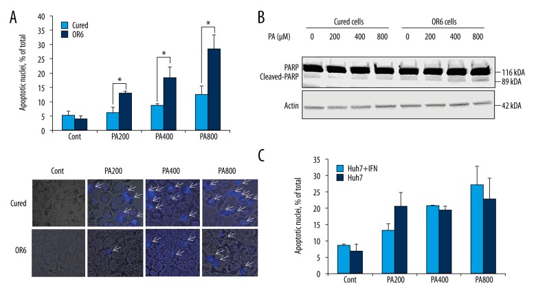
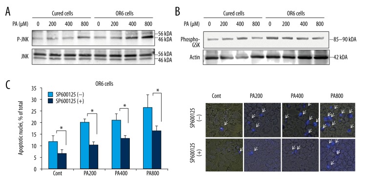
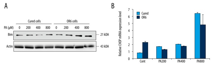
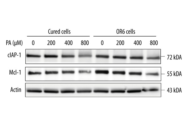
BACKGROUND Hepatitis C virus (HCV) infection and metabolic diseases including nonalcoholic steatohepatitis (NASH) exhibit a complex interplay. Although free fatty acid-mediated apoptosis is a prominent feature of NASH, the impact of HCV infection on hepatocyte lipotoxicity has remained largely unexplored. The study aimed at identifying whether infection by HCV affected the apoptotic pathway in hepatocytes during fatty acid assault. MATERIAL AND METHODS OR6 cells, which are derived from human hepatocellular carcinoma Huh-7 cells and harbor a full-length HCV RNA genome replication system, were treated with palmitate. Apoptosis was examined by 4',6-diamidino-2-phenylindole staining. Activation and expression of JNK, Bim, cIAP-1, and Mcl-1 were examined by immunoblotting. mRNA expression of CHOP, a major player in endoplasmic reticulum stress-mediated apoptosis, was assessed by real-time PCR. RESULTS Palmitate-induced hepatocyte apoptosis was significantly enhanced in OR6 cells compared to cured cells, in which the HCV genome had been eradicated by treatment with interferon-α. Although basal expression of CHOP mRNA was enhanced in OR6 cells compared to cured cells, it was similarly upregulated in both cell lines following palmitate treatment. Notably, palmitate-induced JNK phosphorylation was accentuated in OR6 cells compared to cured cells. Inhibition of JNK with SP600125 attenuated palmitate-induced apoptosis. Palmitate-mediated upregulation of BH3-only protein Bim, which acts downstream of JNK, was also enhanced in OR6 cells compared to cured cells. In contrast, Mcl-1 and cIAP-1 were equally reduced in OR6 cells and cured cells following palmitate treatment. CONCLUSIONS These findings suggest that during lipoapoptosis, HCV infection may enhance hepatocyte toxicity by increasing JNK phosphorylation.
Conflict of interest statement
The authors have no conflict of interest to declare.
Figures
Similar articles
-
Palmitoleate attenuates palmitate-induced Bim and PUMA up-regulation and hepatocyte lipoapoptosis.J Hepatol. 2010 Apr;52(4):586-93. doi: 10.1016/j.jhep.2010.01.003. Epub 2010 Feb 13. J Hepatol. 2010. PMID: 20206402 Free PMC article.
-
Mechanisms of lysophosphatidylcholine-induced hepatocyte lipoapoptosis.Am J Physiol Gastrointest Liver Physiol. 2012 Jan 1;302(1):G77-84. doi: 10.1152/ajpgi.00301.2011. Epub 2011 Oct 13. Am J Physiol Gastrointest Liver Physiol. 2012. PMID: 21995961 Free PMC article.
-
Saturated free fatty acid sodium palmitate-induced lipoapoptosis by targeting glycogen synthase kinase-3β activation in human liver cells.Dig Dis Sci. 2014 Feb;59(2):346-57. doi: 10.1007/s10620-013-2896-2. Epub 2013 Oct 17. Dig Dis Sci. 2014. PMID: 24132507
-
Apoptosis and non-alcoholic fatty liver diseases.World J Gastroenterol. 2018 Jul 7;24(25):2661-2672. doi: 10.3748/wjg.v24.i25.2661. World J Gastroenterol. 2018. PMID: 29991872 Free PMC article. Review.
-
Molecular mechanisms of lipotoxicity and glucotoxicity in nonalcoholic fatty liver disease.Metabolism. 2016 Aug;65(8):1049-61. doi: 10.1016/j.metabol.2016.02.014. Epub 2016 Mar 3. Metabolism. 2016. PMID: 26997538 Free PMC article. Review.
Cited by
-
Function of Mitogen-Activated Protein Kinases in Hepatic Inflammation.J Cell Signal. 2021;2(3):172-180. J Cell Signal. 2021. PMID: 34557866 Free PMC article.
-
Receptors for Respiratory Syncytial Virus Infection and Host Factors Regulating the Life Cycle of Respiratory Syncytial Virus.Front Cell Infect Microbiol. 2022 Feb 25;12:858629. doi: 10.3389/fcimb.2022.858629. eCollection 2022. Front Cell Infect Microbiol. 2022. PMID: 35281439 Free PMC article. Review.
-
Endoplasmic Reticulum Stress in Hepatitis B Virus and Hepatitis C Virus Infection.Viruses. 2022 Nov 25;14(12):2630. doi: 10.3390/v14122630. Viruses. 2022. PMID: 36560634 Free PMC article. Review.
-
Five-Aminolevulinic Acid (5-ALA) Induces Heme Oxygenase-1 and Ameliorates Palmitic Acid-Induced Endoplasmic Reticulum Stress in Renal Tubules.Int J Mol Sci. 2023 Jun 15;24(12):10151. doi: 10.3390/ijms241210151. Int J Mol Sci. 2023. PMID: 37373300 Free PMC article.
References
-
- Satapathy SK, Sanyal AJ. Epidemiology and natural history of nonalcoholic fatty liver disease. Semin Liver Dis. 2015;35:221–35. - PubMed
-
- Akazawa Y, Nakao K. Lipotoxicity pathways intersect in hepatocytes: Endoplasmic reticulum stress, c-Jun N-terminal kinase-1, and death receptors. Hepatol Res. 2016;46(10):977–84. - PubMed
-
- Barreyro FJ, Kobayashi S, Bronk SF, et al. Transcriptional regulation of Bim by FoxO3A mediates hepatocyte lipoapoptosis. J Biol Chem. 2007;282:27141–54. - PubMed
MeSH terms
Substances
LinkOut - more resources
Full Text Sources
Medical
Research Materials
Miscellaneous
