. 2017 Sep 20;8(1):626.
doi: 10.1038/s41467-017-00729-8.

Bacterial outer membrane vesicles suppress tumor by interferon-γ-mediated antitumor response

Affiliations

Bacterial outer membrane vesicles suppress tumor by interferon-γ-mediated antitumor response

Oh Youn Kim et al. Nat Commun. .

Abstract

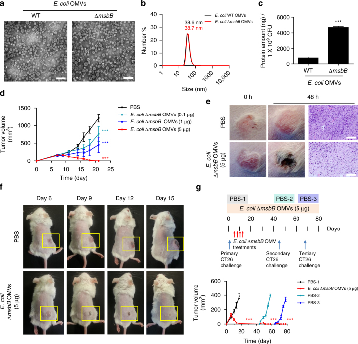
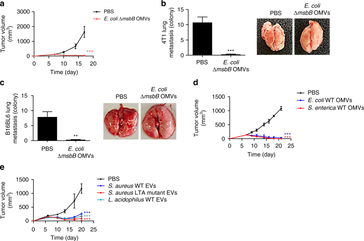
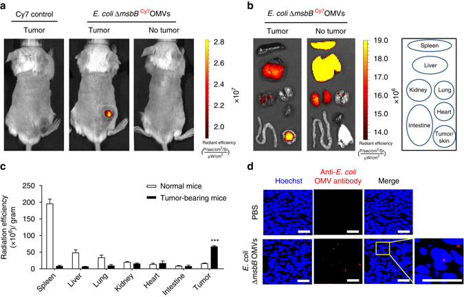
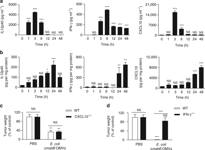
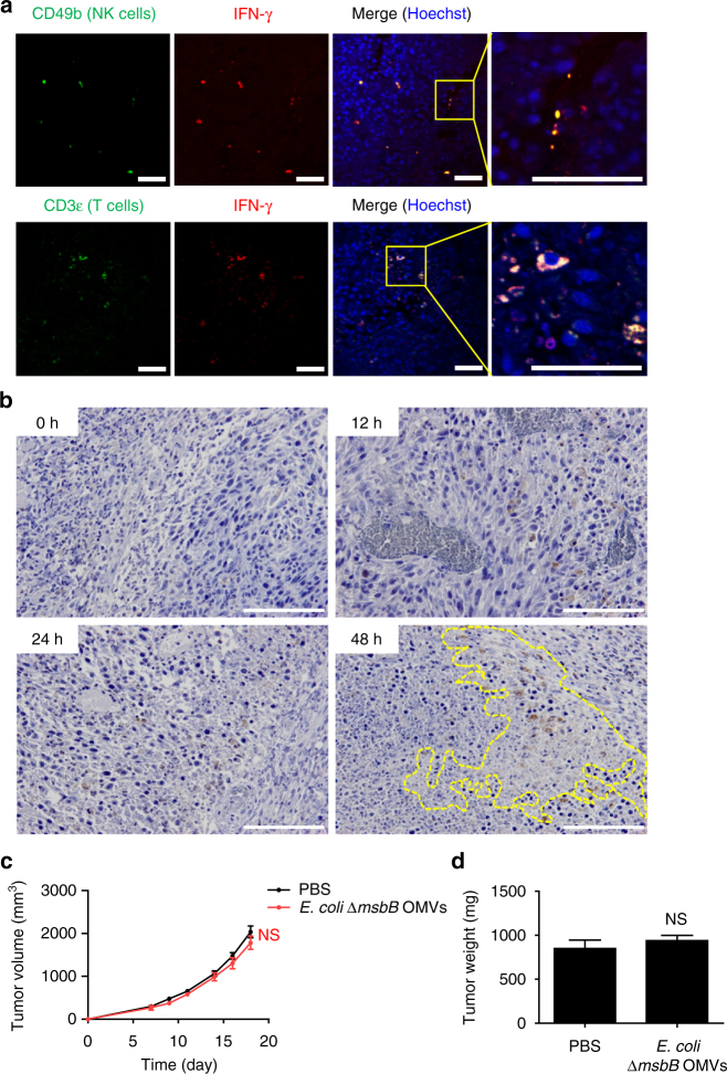
Gram-negative bacteria actively secrete outer membrane vesicles, spherical nano-meter-sized proteolipids enriched with outer membrane proteins, to the surroundings. Outer membrane vesicles have gained wide interests as non-living complex vaccines or delivery vehicles. However, no study has used outer membrane vesicles in treating cancer thus far. Here we investigate the potential of bacterial outer membrane vesicles as therapeutic agents to treat cancer via immunotherapy. Our results show remarkable capability of bacterial outer membrane vesicles to effectively induce long-term antitumor immune responses that can fully eradicate established tumors without notable adverse effects. Moreover, systematically administered bacterial outer membrane vesicles specifically target and accumulate in the tumor tissue, and subsequently induce the production of antitumor cytokines CXCL10 and interferon-γ. This antitumor effect is interferon-γ dependent, as interferon-γ-deficient mice could not induce such outer membrane vesicle-mediated immune response. Together, our results herein demonstrate the potential of bacterial outer membrane vesicles as effective immunotherapeutic agent that can treat various cancers without apparent adverse effects.Bacterial outer membrane vesicles (OMVs) contain immunogens but no study has yet examined their potential in treating cancer. Here, the authors demonstrate that OMVs can suppress established tumours and prevent tumour metastasis by an interferon-γ mediated antitumor response.


Conflict of interest statement

O.Y.K. and Y.S.G. are co-owners of patent entitled ‘Method for treating and diagnosing cancer by using cell-derived microvesicles’ patent number: WO2012002760 A2. The remaining authors declare no competing financial interests.

Figures

Fig. 1
Fig. 1
Treatment of outer membrane vesicles (OMVs) induces complete regression of tumors. a Transmission electron micrograph image of E. coli W3110 wild type-derived OMVs (WT OMVs) and E. coli W3110 msbB mutant-derived OMVs (∆msbB OMVs). Scale bars, 100 nm. b Size distribution of E. coli W3110 WT and ∆msbB OMVs measured by dynamic light scattering analysis (n = 5). c Production yield of E. coli W3110 WT and ∆msbB OMVs from 1 × 109 CFUs bacteria in terms of total protein amount (n = 5, three independent experiments). d Tumor volume of mice bearing CT26 murine colon adenocarcinoma measured after E. colimsbB OMV treatments with various amounts (total n = 14 mice per group, two independent experiments). E. coli ∆msbB OMVs were injected intravenously four times from day 6 with 3 days intervals. e Picture of tumor and tumor tissue histology 48 h after single PBS or ∆msbB OMV (5 μg in total protein amount) injection. Scale bars, 50 μm. f Picture of mice bearing tumor after PBS or ∆msbB OMV (5 μg in total protein amount) treatments. Yellow box indicates tumor sites. g Mice treated with OMV (5 μg in total protein amount) with complete tumor regression were re-challenged with tumors on the opposite flank of the mice 4 weeks after the final injection. Then, these mice were re-challenged with tumor on the middle of the two flanks of the mice 3 weeks after the secondary tumor injection (total n = 14 mice per group, two independent experiments). Data are presented as the mean ± SD from a representative experiments. ***P < 0.001 analysed by unpaired Student’s t-test c or two-way ANOVA with Bonferroni post test to compare each treated group with PBS group d, g
Fig. 2
Fig. 2
Systemic administration of bacterial extracellular vesicles induces effective antitumor responses in multiple tumors. a Tumor volume of mice measured after the subcutaneous injection of MC38 murine adenocarcinoma (total n = 14 mice per group, two independent experiments). E. colimsbB OMVs (5 μg in total protein amount) were injected intravenously four times from day 7 with 3 days intervals. b, c To induce spontaneous lung metastasis, highly metastatic 4T1 murine carcinoma cells and B16BL6 melanoma cells were subcutaneously injected to the right flank of the mice (total n = 12 mice per group, two independent experiments). PBS or E. colimsbB OMVs (5 μg in total protein amount) were intravenously treated for four times with 3 days intervals from day 7. At day 22, mice were killed and the number of colonies spontaneously metastasized in the lungs for 4T1 b and B16BL6 c tumors were counted. d, e Tumor volume of mice bearing CT26 tumor of mice intravenously injected with four doses of 5 μg of Gram-negative E. coli W3110 wild type- and Salmonella enterica wild type-derived OMVs. d Gram-positive bacteria Staphylococcus aureus wild type- and lipoteichoic acid (LTA) mutant-derived extracellular vesicles (EVs), and Lactobacillus acidophilus wild type-derived extracellular vesicles (EVs), e (n = 5 mice per group). Data are presented as the mean ± SD from a representative experiments. *P < 0.01 and **P < 0.001, respectively, analysed by unpaired Student’s t-test b, c or two-way ANOVA a, d, e. Bonferroni post test was applied to compare each treated group with PBS group d, e
Fig. 3
Fig. 3
Targeting of E. coli ∆msbB OMVs to tumor tissues after systemic administration. a Cy7 control and Cy7-labeled E. coli ∆msbB OMVs (∆msbB Cy7OMVs) were systematically injected to BALB/c mice bearing CT26 tumor cells. For control, ∆msbB Cy7OMVs were also injected to healthy BALB/c mice with no tumor. Whole body distributions of the injected Cy7OMVs were observed using in vivo imaging system spectrum 12 h after the injection. b Spleen, liver, kidney, lung, heart, intestine, and tumor tissues were isolated to measure the accumulation of Cy7 fluorescence in different organs. c Radiant efficiencies of each organ were acquired for Cy7 fluorescence using Living Image 3.1 Software and were normalized by each organ weight. Results are from three independent experiments (total n = 9). d For tumor tissue immunohistochemistry, ∆msbB OMVs were intravenously injected to BALB/c mice bearing CT26 tumor. Tumor tissues were extracted after 12 h and were embedded in paraffin for tissue section analyses by immunohistochemistry. The cell nucleus is stained in blue (Hoechst) and OMVs are shown in red fluorescence signal (anti-E. coli OMV polyclonal antibodies) Scale bars, 50 μm. ***P < 0.001 analysed by unpaired Student’s t-test
Fig. 4
Fig. 4
E. coli ∆msbB OMV antitumor effect is IFN-γ dependent. a, b Release of antitumor cytokines IL-12p40, IFN-γ, and CXCL10 in blood sera a and tumor cell lysate b after single intravenous injection of E. colimsbB OMVs (5 μg in total protein amount) to mice bearing CT26 tumors at different time points. c, d The antitumor efficacy of OMV treatment in CXCL10-deficient (CXCL10−/−) mice c and IFN-γ-deficient (IFN-γ−/−) mice d. Data are shown as the mean ± SD (n = 6 mice per group). NS, not significant, *P < 0.05, **P < 0.01, and ***P < 0.001, respectively, analysed by one-way ANOVA. Bonferroni multiple comparisons post test was applied to compare each time point to zero time point a, b or to compare treated group with PBS group of each mouse c, d
Fig. 5
Fig. 5
Importance of NK and T cells on OMV antitumor effect. a Images of tumor tissues isolated from wild-type mice bearing CT26 tumors, stained for IFN-γ, and NK cells (top) or T cells (bottom) 48 h after intravenous injections of E. colimsbB OMVs. The cell nucleus is stained in blue (Hoechst), whereas NK and T cells are shown by green fluorescence signal and IFN-γ is shown in red fluorescence signal, respectively. Scale bars, 50 μm. b Images of tumor tissues isolated from wild-type mice bearing CT26 tumors, stained for NK cells (brown spots) at different time points after intravenous injections of E. colimsbB OMVs. Tumor necrotic area around NK cells at 48 h is shown in dashed lines. Scale bars, 50 μm. c, d Tumor volume of NIHS-LystbgFoxn1nuBtkxid mice bearing CT26 tumor measured after E. colimsbB OMV treatments c and tumor weight at the end of the experiment d. Data are presented as the mean ± SD (n = 6 mice per group). NS indicates not significant analysed by two-way ANOVA c and unpaired Student’s t-test d

References

    1. Sharma P, Allison JP. The future of immune checkpoint therapy. Science. 2015;348:56–61. doi: 10.1126/science.aaa8172. - DOI - PubMed
    1. Fesnak AD, June CH, Levine BL. Engineered T cells: the promise and challenges of cancer immunotherapy. Nat. Rev. Cancer. 2016;16:566–581. doi: 10.1038/nrc.2016.97. - DOI - PMC - PubMed
    1. Sharma P, Allison JP. Immune checkpoint targeting in cancer therapy: toward combination strategies with curative potential. Cell. 2015;161:205–214. doi: 10.1016/j.cell.2015.03.030. - DOI - PMC - PubMed
    1. Pardoll DM. The blockade of immune checkpoints in cancer immunotherapy. Nat. Rev. Cancer. 2012;12:252–264. doi: 10.1038/nrc3239. - DOI - PMC - PubMed
    1. Kershaw MH, Westwood JA, Darcy PK. Gene-engineered T cells for cancer therapy. Nat. Rev. Cancer. 2013;13:525–541. doi: 10.1038/nrc3565. - DOI - PubMed

Publication types

MeSH terms