. 2017 Sep 20;7(1):11988.
doi: 10.1038/s41598-017-12154-4.

THP-1-derived macrophages render lung epithelial cells hypo-responsive to Legionella pneumophila - a systems biology study

Affiliations

THP-1-derived macrophages render lung epithelial cells hypo-responsive to Legionella pneumophila - a systems biology study

Christine Schulz et al. Sci Rep. .

Abstract

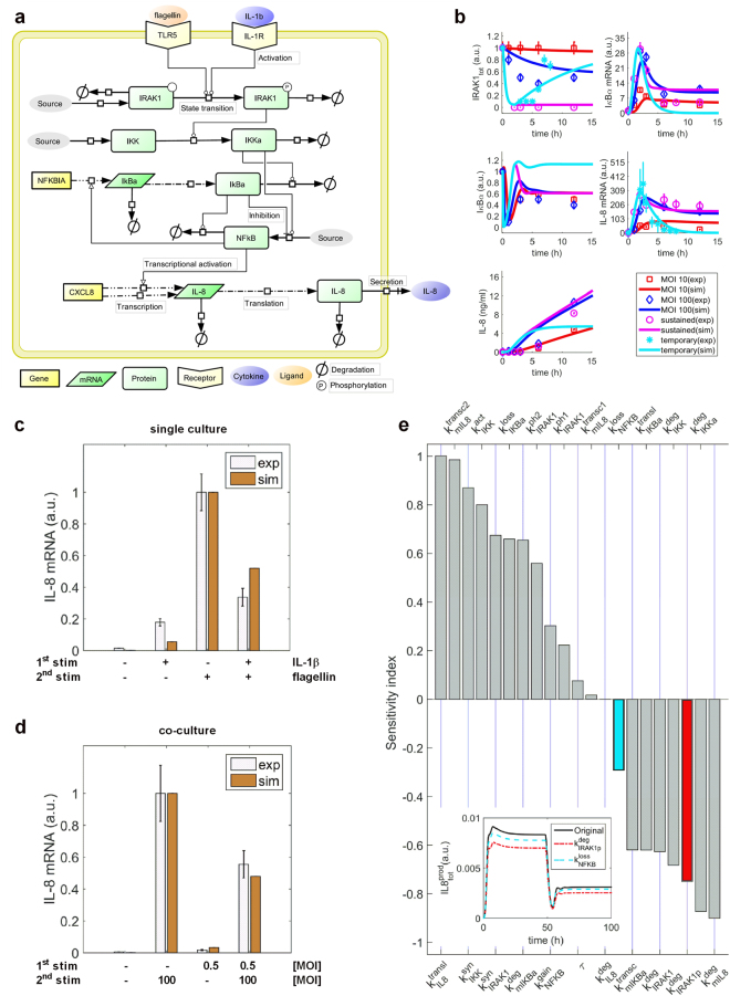
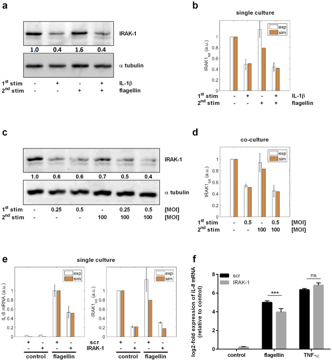
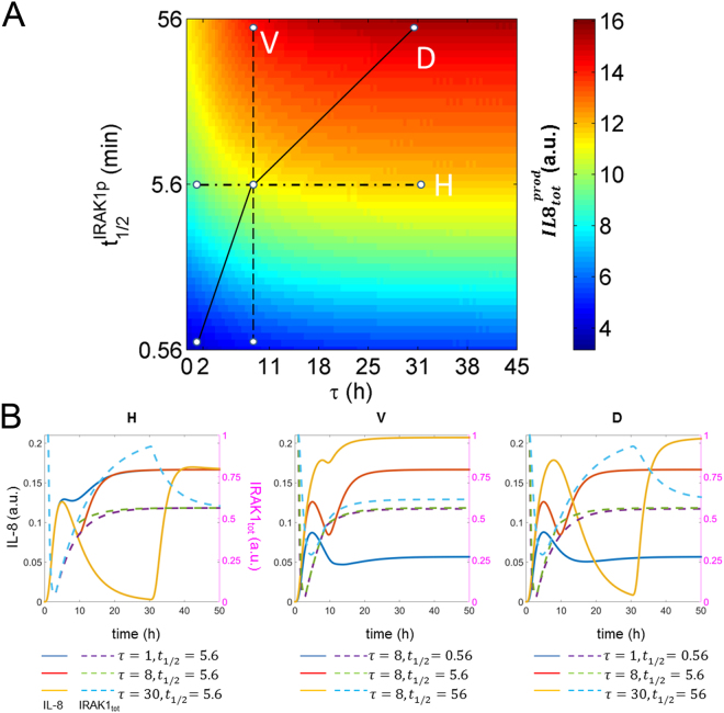
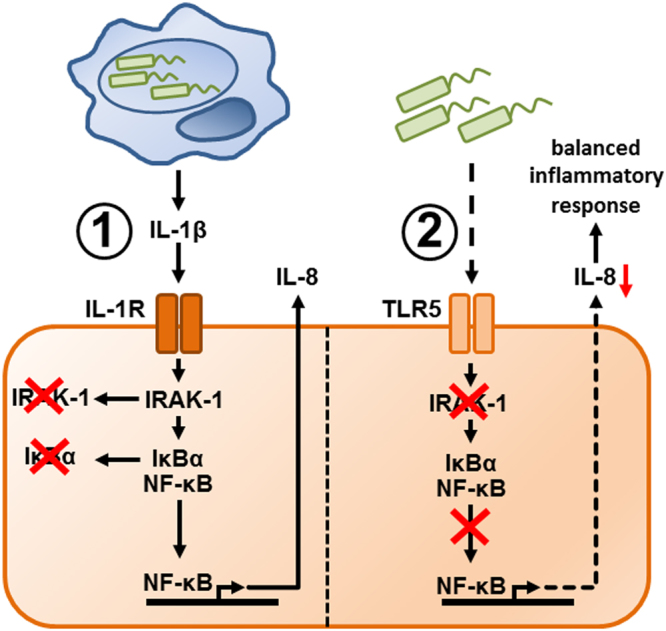
Immune response in the lung has to protect the huge alveolar surface against pathogens while securing the delicate lung structure. Macrophages and alveolar epithelial cells constitute the first line of defense and together orchestrate the initial steps of host defense. In this study, we analysed the influence of macrophages on type II alveolar epithelial cells during Legionella pneumophila-infection by a systems biology approach combining experimental work and mathematical modelling. We found that L. pneumophila-infected THP-1-derived macrophages provoke a pro-inflammatory activation of neighboring lung epithelial cells, but in addition render them hypo-responsive to direct infection with the same pathogen. We generated a kinetic mathematical model of macrophage activation and identified a paracrine mechanism of macrophage-secreted IL-1β inducing a prolonged IRAK-1 degradation in lung epithelial cells. This intercellular crosstalk may help to avoid an overwhelming inflammatory response by preventing excessive local secretion of pro-inflammatory cytokines and thereby negatively regulating the recruitment of immune cells to the site of infection. This suggests an important but ambivalent immunomodulatory role of macrophages in lung infection.


Conflict of interest statement

The authors declare that they have no competing interests.

Figures

Figure 1
Figure 1
Infection of THP-1 cells with L. pneumophila induces pro-inflammatory cytokine release. THP-1 cells were stimulated with L. pneumophila (MOI 0.5) for 24 or 48 h, respectively. (a) IL-8 secretion was measured by ELISA, (be) secretion of IL-1β, TNF-α, IL-6 and IL-10 were measured using Multiplex Luminex Assay. Data are shown as mean ± SEM (n = 4). 2-Way ANOVA was performed with Sidak’s multiple comparison test as described: *Compared to corresponding control, #compared to equally treated 24 h sample; #or *p ≤ 0.05, ##p ≤ 0.01, ###or ***p ≤ 0.001, ****or ####p ≤ 0.0001.
Figure 2
Figure 2
Infection of THP-1 cells with L. pneumophila induces paracrine activation of co-cultured lung epithelial cells. (a,b) THP-1 cells were stimulated with L. pneumophila (MOI 0.5) for 24 or 48 h. (A) IL-8 secretion of co-cultures was measured by ELISA; black line: cytokine release of THP-1 cells 48 h post infection calculated according to Fig. 1. (b) IL-8 expression of co-cultured A549 cells was analysed by RT-qPCR. (a,b) Data are shown as mean ± SEM (n = 4). (c,d) Co-cultures were pre-incubated either with (c) IL-1ra (50 or 100 ng/mL) or (d) anti-TNF-α (7.5 or 15 ng/mL) together with 100 ng/mL IL-1ra for 1 h. Subsequently, THP-1 cells were stimulated with L. pneumophila (MOI 0.5) for 48 h. IL-8 expression of A549 cells was analysed by RT-qPCR. Data are shown as mean ± SEM (n = 3). 2-Way ANOVA was performed with Sidak’s multiple comparison test as described: *Compared to corresponding control, #compared to equally treated 24 h sample, §compared to matching L. pneumophila-infected sample; #p ≤ 0.05, ***or ###or §§§p ≤ 0.001, ****or §§§§p ≤ 0.0001, ns = not significant.
Figure 3
Figure 3
Paracrine mechanisms render alveolar epithelial cells hypo-responsive to L. pneumophila infection. (ac) THP-1 cells were stimulated with L. pneumophila (MOI 0.5) for 48 h. Upon their removal, A549 cells were incubated in fresh medium (4 h) followed by L. pneumophila infection (MOI 100) for 3 h. Expression of (a,c) IL-8 or (b) IL-6 was analysed by RT-qPCR. (c) Co-cultures were stimulated with IL-1ra (100 ng/mL) for 1 h prior to re-stimulation. (ac) Data are shown as average percentage ± SEM of individual pro-inflammatory mRNA induction relative to L. pneumophila (MOI 100) treatment (n = 4). (d) A549 cells were stimulated with IL-1β (1 ng/mL) for 2 h. Following medium renewal (4 h incubation), A549 cells were stimulated with flagellin (100 ng/mL) for 3 h. IL-8 expression was analysed by RT-qPCR. Data are shown as mean ± SEM (n = 4). Two-tailed Student’s t-test (a,b,d) or 2-Way ANOVA with Sidak’s multiple comparison test (c) were performed as described: (ac) *compared to L. pneumophila-infected sample (MOI 100), #compared to corresponding IL-1ra-untreated sample; *p ≤ 0.05, **p ≤ 0.01, ###p ≤ 0.001, ns = not significant. (d) §Compared to flagellin stimulation; §§§p ≤ 0.001.
Figure 4
Figure 4
Hypo-responsiveness of lung epithelial cells is accompanied by changes in RNA half-life and IκBα-regulated nuclear presence of p65. (a) A549 cells were incubated with TSA (50 nM) or Parg (3 µM) for 2 h prior to stimulation with IL-1β (1 ng/mL, 2 h), medium renewal (4 h) and flagellin stimulation (100 ng/mL, 3 h). IL-8 expression was analysed by RT-qPCR. Data are shown as mean ± SEM (n = 3). (be) A549 cells were stimulated with IL-1β (1 ng/mL, 2 h) followed by medium renewal. (b,c) Then, A549 cells were incubated with actinomycin D (10 µM, up to 360 min). (b) IL-8 expression was analysed by RT-qPCR. (c) Data from B are shown as mean percentage ± SEM of individual IL-8 induction relative to IL-1β stimulation (n = 4). IL-8 half-life was calculated using non-linear regression. (d,e) After 4 h of incubation, A549 cells were stimulated with flagellin (100 ng/mL, 30 min). Nuclear and cytosolic presence of p65, α tubulin, and lamin C (d) and protein levels of IκBα and α tubulin (e) were analysed by western blotting and quantified. One out of three representative blots is shown. 2-Way ANOVA was performed with Sidak’s multiple comparison test as described: (a) *Compared to corresponding flagellin-stimulated sample, #compared to corresponding untreated sample; #p ≤ 0.05, ****p ≤ 0.001, ns = not significant.
Figure 5
Figure 5
Mathematical modelling of the NF-κB signalling pathway. (a) Model scheme. (b) Model calibration. The model was fitted using time-series data: sustained challenge of lung epithelial cells with L. pneumophila (MOI 10 or MOI 100, up to 24 h); A549 cell stimulation with IL-1β (1 ng/mL) for up to 12 h (sustained) or for 2 h followed by medium renewal and further incubation for up to 24 h (temporary). Lines represent model simulations and the symbols account for experimental data (mean ± SD). (c,d) Model validation. The data-driven model possesses the ability to predict experimental data (mean ± SEM) showing the reduced IL-8 mRNA expression of lung epithelial cells after pre-stimulation. (c) A549 cells were stimulated with IL-1β (1 ng/mL, 2 h) followed by medium renewal (4 h incubation) and flagellin stimulation (100 ng/mL, 3 h). (d) THP-1 cells were stimulated with L. pneumophila (MOI 0.5) for 48 h. Upon their removal, A549 cells were incubated in fresh medium (4 h) followed by L. pneumophila infection (MOI 100) for 3 h. (e) Sensitivity analysis. kIRAK1deg and kIRAK1pdeg rank in top five parameters to negatively influence IL-8 production. For illustration, the inset figure shows the influence of kIRAK1pdeg (red) and kNFkBloss (cyan) on IL-8 production after increasing their original values by 25%. kIRAK1pdeg has stronger impact on reduction of IL-8 production. a.u.: arbitrary unit. IL-1R: IL-1 receptor.
Figure 6
Figure 6
Hypo-responsiveness is mediated by IRAK-1 and can be mimicked by its knockdown. (a,b) A549 cells were stimulated with IL-1β (1 ng/mL, 2 h) followed by medium renewal (4 h incubation) and flagellin stimulation (100 ng/mL, 3 h). (a) Protein expression was analysed by western blotting and quantified. One out of four representative blots is shown. (b) The experimental data were used to validate model predictions. (c,d) THP-1 cells were stimulated with L. pneumophila (MOI 0.25 or 0.5) for 48 h. Upon their removal and subsequent medium renewal (4 h incubation), A549 cells were stimulated with L. pneumophila (MOI 100, 3 h). (c) Protein levels of IRAK-1 and α tubulin were analysed by western blotting and quantified. One out of four representative blots is shown. (d) Model predictions were validated using experimental data. (e,f) A549 cells were transfected with control (scr) or IRAK-1 siRNA (48 h) and stimulated with flagellin (100 ng/mL) or TNF-α (100 ng/mL) for 3 h. (e) The model was used to predict IL-8 expression upon IRAK-1 depletion, which was in accordance with the experimental data. (f) IL-8 expression was analysed by RT-qPCR and normalized to an untreated scr control (mean ± SEM, n = 4). (e) The data were compared with model predictions. Statistical analysis: (f) 2-Way ANOVA with Sidak’s multiple comparison test as described; *Compared to corresponding scr, ***p ≤ 0.001; ns = not significant.
Figure 7
Figure 7
In silico modelling of lung epithelial cell hypo-responsiveness to re-stimulation. (A) Overall production of IL-8 (IL8totprod) for different combined values of τ and t1/2IRAK1p is shown. τ represents the interval between the two stimuli: IL-1β and flagellin. t1/2IRAK1p denotes for the half-life of phosphorylated IRAK-1, and its wild type value is 5.6 min. (B) Temporal dynamics of IL-8 (IL8; solid lines) and total amounts of IRAK-1 (IRAK1 tot; dashed lines) are presented for different combinations of τ and t1/2IRAK1p. The selected combinations of τ and t1/2IRAK1p (H,V and D) are delineated by different types of lines in a.
Figure 8
Figure 8
Macrophages can induce hypo-responsiveness of epithelial cells to L. pneumophila infection through a paracrine mechanism. Macrophages (blue) induce tolerance of neighboring lung epithelial cells (orange) to L. pneumophila (green) infection through an IL-1β-dependent mechanism (①), which results in prolonged depletion of IRAK-1 in the epithelial cells (②). This mechanism attenuates subsequent pro-inflammatory activation of these epithelial cells upon TLR5-dependent recognition of L. pneumophila and thus prevents potentially harmful inflammatory reactions in the lung.

Similar articles

Cited by

References

    1. Armstrong GL, Conn LA, Pinner RW. Trends in infectious disease mortality in the United States during the 20th century. JAMA. 1999;281:61–66. doi: 10.1001/jama.281.1.61. - DOI - PubMed
    1. Mizgerd JP. Lung infection–a public health priority. PLoS Med. 2006;3:e76. doi: 10.1371/journal.pmed.0030076. - DOI - PMC - PubMed
    1. Welte T, Torres A, Nathwani D. Clinical and economic burden of community-acquired pneumonia among adults in Europe. Thorax. 2012;67:71–79. doi: 10.1136/thx.2009.129502. - DOI - PubMed
    1. WHO. Pneumonia (2015).
    1. Wardlaw, T., White Johansson, E. & Hodge M. Pneumonia: The forgotten killer of children. (UNICEF/WHO 2006).

Publication types