

Accuracy and Reliability of Cone-Beam Computed Tomography for Linear and Volumetric Mandibular Condyle Measurements. A Human Cadaver Study
- PMID: 28931867
- PMCID: PMC5607232
- DOI: 10.1038/s41598-017-12100-4
Accuracy and Reliability of Cone-Beam Computed Tomography for Linear and Volumetric Mandibular Condyle Measurements. A Human Cadaver Study
Abstract
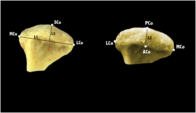
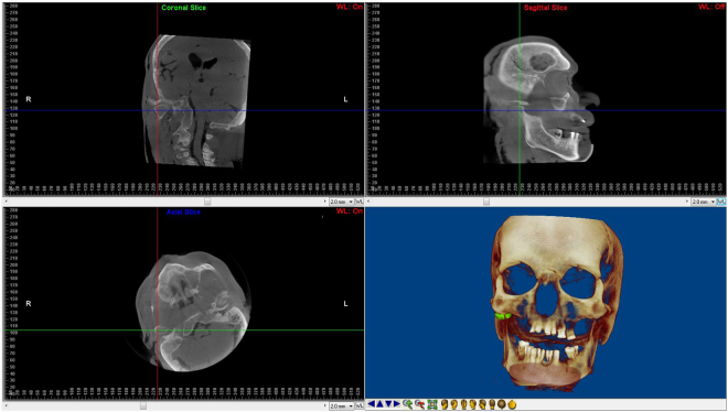
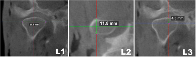
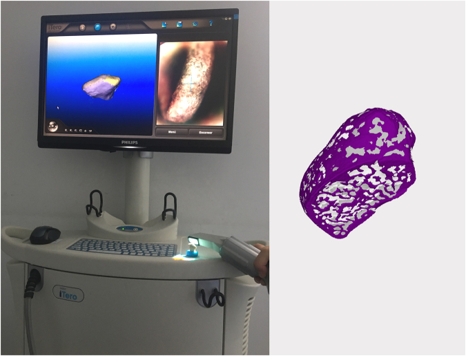
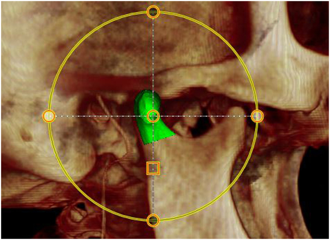
The accuracy of Cone-Beam Computed Tomography (CBCT) on linear and volumetric measurements on condyles has only been assessed on dry skulls. The aim of this study was to evaluate the reliability and accuracy of linear and volumetric measurements of mandibular condyles in the presence of soft tissues using CBCT. Six embalmed cadaver heads were used. CBCT scans were taken, followed by the extraction of the condyles. The water displacement technique was used to calculate the volumes of the condyles and three linear measurements were made using a digital caliper, these measurements serving as the gold standard. Surface models of the condyles were obtained using a 3D scanner, and superimposed onto the CBCT images. Condyles were isolated on the CBCT render volume using the surface models as reference and volumes were measured. Linear measurements were made on CBCT slices. The CBCT method was found to be reliable for both volumetric and linear measurements (CV < 3%; CCI > 0.90). Highly accurate values were obtained for the three linear measurements and volume. CBCT is a reliable and accurate method for taking volumetric and linear measurements on mandibular condyles in the presence of soft tissue, and so a valid tool for clinical diagnosis.
Conflict of interest statement
The authors declare that they have no competing interests.
Figures

Similar articles
-
Cone beam computed tomography 3D reconstruction of the mandibular condyle.Angle Orthod. 2008 Sep;78(5):880-8. doi: 10.2319/072007-339.1. Angle Orthod. 2008. PMID: 18298200
-
Volumetric analysis of the mandibular condyle using cone beam computed tomography.Eur J Radiol. 2012 Aug;81(8):1812-6. doi: 10.1016/j.ejrad.2011.04.070. Epub 2011 Jun 15. Eur J Radiol. 2012. PMID: 21680124
-
Validation of a novel semi-automated method for three-dimensional surface rendering of condyles using cone beam computed tomography data.Int J Oral Maxillofac Surg. 2013 Aug;42(8):1023-9. doi: 10.1016/j.ijom.2013.01.016. Epub 2013 Mar 23. Int J Oral Maxillofac Surg. 2013. PMID: 23528746
-
Reliability and accuracy of segmentation of mandibular condyles from different three-dimensional imaging modalities: a systematic review.Dentomaxillofac Radiol. 2020 Jul;49(5):20190150. doi: 10.1259/dmfr.20190150. Epub 2019 Dec 3. Dentomaxillofac Radiol. 2020. PMID: 31778321 Free PMC article.
-
Cone-beam computed tomography imaging of dentoalveolar and mandibular fractures.Oral Radiol. 2020 Jul;36(3):217-224. doi: 10.1007/s11282-019-00390-5. Epub 2019 May 17. Oral Radiol. 2020. PMID: 31102106 Review.
Cited by
-
Three dimensional condylar positional and morphological changes following mandibular reconstruction based on CBCT analysis: a prospective study.Head Face Med. 2023 Feb 7;19(1):3. doi: 10.1186/s13005-023-00347-4. Head Face Med. 2023. PMID: 36747208 Free PMC article.
-
Evaluation of associations between condylar morphology, ramus height, and mandibular plane angle in various vertical skeletal patterns: a digital radiographic study.BMC Oral Health. 2022 Aug 8;22(1):330. doi: 10.1186/s12903-022-02365-1. BMC Oral Health. 2022. PMID: 35941596 Free PMC article.
-
Evaluation of the linear and volumetric measuring changes in different positions in CBCT.Clin Exp Dent Res. 2023 Jun;9(3):535-542. doi: 10.1002/cre2.732. Epub 2023 Apr 4. Clin Exp Dent Res. 2023. PMID: 37016557 Free PMC article.
-
Accuracy of facial skeletal surfaces segmented from CT and CBCT radiographs.Sci Rep. 2023 Nov 28;13(1):21002. doi: 10.1038/s41598-023-48320-0. Sci Rep. 2023. PMID: 38017262 Free PMC article.
-
Appraisal of the Accuracy and Reliability of Cone-Beam Computed Tomography and Three-Dimensional Printing for Volumetric Mandibular Condyle Measurements of a Human Condyle.Cureus. 2023 Oct 9;15(10):e46746. doi: 10.7759/cureus.46746. eCollection 2023 Oct. Cureus. 2023. PMID: 38022326 Free PMC article.
References
-
- Veyre-Goulet S, Fortin T, Thierry A. Accuracy of linear measurement provided by cone beam computed tomography to assess bone quantity in the posterior maxilla: a human cadaver study. Clin Implant Dent Relat Res. 2008;10:226–30. - PubMed
Publication types
MeSH terms
LinkOut - more resources
Full Text Sources
Other Literature Sources
