. 2017 Sep 20;7(1):11992.
doi: 10.1038/s41598-017-12269-8.

Netrin-1 Derived from the Ventricular Zone, but not the Floor Plate, Directs Hindbrain Commissural Axons to the Ventral Midline

Affiliations

Netrin-1 Derived from the Ventricular Zone, but not the Floor Plate, Directs Hindbrain Commissural Axons to the Ventral Midline

Kenta Yamauchi et al. Sci Rep. .

Abstract

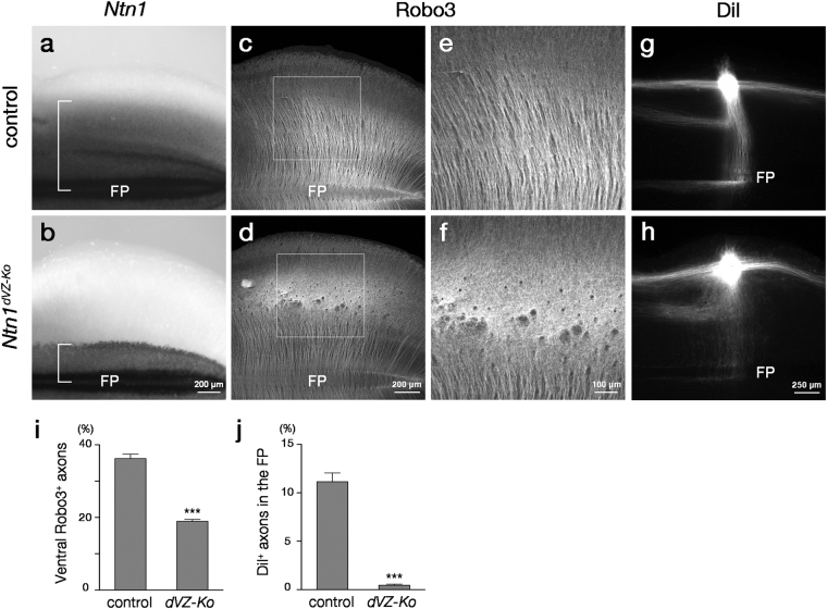
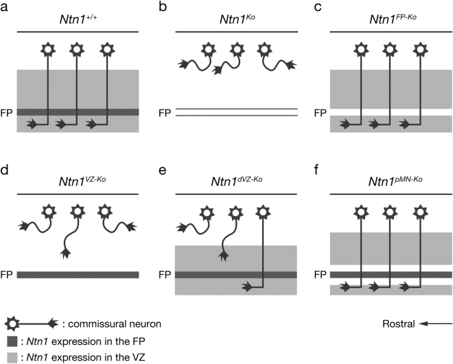
Netrin-1 (Ntn1) emanating from the ventral midline has been thought to act as a long-range diffusible chemoattractant for commissural axons (CAs). However, CAs still grow towards the midline in the absence of the floor plate (FP), a glial structure occupying the midline. Here, using genetically loss-of-function approaches in mice, we show that Ntn1 derived from the ventricular zone (VZ), but not the FP, is crucial for CA guidance in the mouse hindbrain. During the period of CA growth, Ntn1 is expressed in the ventral two-thirds of the VZ, in addition to the FP. Remarkably, deletion of Ntn1 from the VZ and even from the dorsal VZ highly disrupts CA guidance to the midline, whereas the deletion from the FP has little impact on it. We also show that the severities of CA guidance defects found in the Ntn1 conditional mutants were irrelevant to their FP long-range chemoattractive activities. Our results are incompatible with the prevailing view that Ntn1 is an FP-derived long-range diffusible chemoattractant for CAs, but suggest a novel mechanism that VZ-derived Ntn1 directs CAs to the ventral midline by its local actions.


Conflict of interest statement

The authors declare that they have no competing interests.

Figures

Figure 1
Figure 1
The spatiotemporal relationship between Ntn1 expression and CA growth in the developing mouse hindbrain. (ac) X-gal staining in whole-mount preparations of E10.5 (a), E11.5 (b) and E12.5 (c) Ntn1 +/LacZ mouse hindbrains (E10.5, n = 12; E11.5, n = 8; E12.5, n = 8). The Ntn1 +/LacZ mouse harbors a ß-geo gene trap vector, allowing Ntn1 expressions to depict by X-gal histochemistry. X-gal reaction products are found in the approximately ventral two-thirds of the hindbrain, in addition to the FP. Dorsal is upwards and rostral is towards the left. (dg) X-gal histochemistry (blue) and Robo3 immunostaining (brown) in E9.5 (d), E10.5 (e) E11.5 (f) and E12.5 (g) Ntn1 +/LacZ mouse hindbrain transverse sections at the rhombomere 7/8 level (E9.5, n = 4, E10.5, n = 4; E11.5, n = 3; E12.5, n = 3). At all these stages, X-gal products are detected in the FP and the ventral two-thirds of VZ along the entire or most part of the circumferential path of Robo3+ axons. Note that X-gal reaction products do not necessarily reflect Ntn1 protein localization. CP, cerebellar primodium; TG, trigeminal ganglia. The bar in (a) and (g) apply to (ac) and (dg), respectively.
Figure 2
Figure 2
Generation of Ntn1 FP-Ko and Ntn1 VZ-Ko mice. (a) Targeting strategy for an Ntn1 conditional allele. Schematic diagram of the Ntn1 locus, targeting vector, targeted allele (Ntn1 FRT-neo), conditional allele (Ntn1 flox) and null allele (Ntn1 ). Ntn1 flox mice were generated by flanking the Ntn1 exon 2 with loxP sites. (b,c) Multiplex PCR genotyping of the targeted allele (b) and the conditional and null allele (c) using primers P1 (Ntn1 Frt Fw5), P2 (Ntn1 Frt Rv3), P3 (Pgk Pr2) and P4 (Ntn1 ∆ Fw2). (dg) Ntn1 ISH in whole-mount preparations of E12.5 Ntn1 +/+ (d), Ntn1 Ko (e), Ntn1 FP-Ko (f) and Ntn1 VZ-Ko (g) mouse hindbrains (Ntn1 +/+, n = 6; Ntn1 Ko, n = 5; Ntn1 FP-Ko, n = 5; Ntn1 VZ-Ko, n = 6). Ntn1 is expressed in the FP and the ventral two-thirds of lateral neural tube in Ntn1 +/+ mice (d). Ntn1 hybridization signals are not detected in Ntn1 Ko mice (e). In Ntn1 FP-Ko mice, Ntn1 expression in the FP is deleted and that in the lateral domain is slightly reduced (f). The laterally expressed Ntn1 is specifically deleted in Ntn1 VZ-ko mice (g). Dorsal is upwards and rostral is towards the left. Hindbrains at the rhombomere 6–8 level are represented.
Figure 3
Figure 3
Aberrant CA growth caused by deletion of Ntn1 from the VZ. (ad) Robo3 immunostaining in whole-mount preparations of E12.5 Ntn1 +/+ (a), Ntn1 Ko (b), Ntn1 FP-Ko (c) and Ntn1 VZ-Ko (d) mouse hindbrains (Ntn1 +/+, n = 7; Ntn1 Ko, n = 5; Ntn1 FP-Ko, n = 8; Ntn1 VZ-Ko; n = 7). Robo3+ axons grow ventrally and reach the FP in Ntn1 +/+ and Ntn1 FP-Ko mice (a,c). Ventrally growing Robo3+ axons were highly disorganized and their number is markedly reduced in Ntn1 Ko and Ntn1 VZ-Ko mice (b,d). (e) Histograms representing the fluorescence intensity of Robo3+ axons within the ventral one-fourth of the hindbrain normalized to that in the preparation (Ntn1 +/+, n = 7; Ntn1 Ko, n = 5; Ntn1 FP-Ko, n = 8; Ntn1 VZ-Ko, n = 7; n.s P > 0.05, *P < 0.05, **P < 0.01; Kruskal-Wallis test followed by Steel post hoc test; P Ko = 0.0126, P FP-Ko = 0.159, P VZ-Ko = 0.00493). Error bars indicate SEM. (f–i) DiI labeling of CAs in whole-mount preparations of E12.5 Ntn1 +/+ (f), Ntn1 ko (g), Ntn1 FP-ko (h) and Ntn1 VZ-ko (i) mouse hindbrains (Ntn1 +/+, n = 6; Ntn1 Ko, n = 9; Ntn1 FP-Ko, n = 9; Ntn1 VZ-Ko, n = 8). DiI crystals are implanted into the dorsal hindbrain at the rhombomere 7/8 level. In Ntn1 +/+ mice, DiI-labeled CAs grow straight towards the FP (arrowheads) and crossed it (f). Longitudinally extending axons in the dorsal margin and ipsilaterally turning axons at the middle along the dorsoventral axis are also normally labeled (f). In Ntn1 Ko mice, ventrally directed axons are foreshortened, spread rostrocaudally and fail to invade ventral hindbrain (g). Ntn1 FP-Ko mice exhibit axon trajectories almost identical to those of Ntn1 +/+ mice (h, arrowheads), whereas Ntn1 VZ-Ko mice exhibit CA guidance defects similar to those of Ntn1 Ko mice (i). (j) Histograms representing the fluorescence intensity of DiI-labeled axons within the FP normalized to that at the implantation site (Ntn1 +/+, n = 6; Ntn1 Ko, n = 9; Ntn1 FP-Ko, n = 9; Ntn1 VZ-Ko, n = 8; n.s P > 0.05, **P < 0.01, ***P < 0.001; Kruskal-Wallis test followed by Steel post hoc test; P Ko = 8.65 × 10−4, P FP-Ko = 0.334, P VZ-Ko = 0.00533). Error bars indicate SEM. Dorsal is upwards and rostral is towards the left. Hindbrains at the rhombomere 6–8 level are represented. The bar in (d) and (i) apply to (ad) and (fi), respectively.
Figure 4
Figure 4
CA guidance defects caused by deletion of Ntn1 from the dorsal VZ. (a,b) Ntn1 ISH in whole-mount preparations of E12.5 control (Pax3 +/Cre ;Ntn1 +/flox) (a) and Ntn1 dVZ-Ko (b) mouse hindbrains (control, n = 3; Ntn1 dVZ-Ko, n = 7). Dorsal expression of Ntn1 is deleted in Ntn1 dVZ-Ko mouse hindbrains. Brackets enclose Ntn1 expression along the dorsoventral axis in each genotype. (c,d) Robo3 immunostaining in whole-mount preparations of E12.5 control (c) and Ntn1 dVZ-Ko (d) mouse hindbrains (control, n = 10; Ntn1 dVZ-Ko, n = 9). (e) and (f) are enlarged views of the dorsal hindbrain regions in (c) and (d), respectively. In Ntn1 dVZ-Ko mice, Robo3+ axons are disorganized in the dorsal hindbrain, reducing the number of ventrally growing Robo3+ axons. (g,h) DiI labeling of CAs in whole-mount preparations of E12.5 control (g) and Ntn1 dVZ-Ko (h) mouse hindbrains (control, n = 9; Ntn1 dVZ-Ko, n = 7). DiI-labeled CAs originated from the dorsal hindbrain are defasciculated and often fail to reach the midline in Ntn1 dVZ-Ko mice (h). (i,j) Histograms representing the fluorescence intensity of Robo3+ axons within the ventral one-fourth of the hindbrain normalized to that in the preparation (control, n = 10; Ntn1 dVZ-Ko, n = 9; ***P < 0.001, Mann–Whitney U-test; P = 2.17 × 10−5) (i) and the fluorescence intensity of DiI-labeled axons within the FP normalized to that at the implantation site (control, n = 9; Ntn1 dVZ-Ko, n = 7; ***P < 0.001, Mann–Whitney U-test; P = 1.75 × 10−4) (j), respectively. Error bars indicate SEM. Dorsal is upwards and rostral is towards the left. Hindbrains at the rhombomere 6–8 level are represented. The bar in (b), (d), (f) and (h) apply to (a,b), (c,d), (e,f) and (g,h), respectively.
Figure 5
Figure 5
Normal CA growth in Ntn1 pMN-Ko mice. (a,b) Ntn1 ISH in whole-mount preparations of E12.5 control (Olig2 +/Cre ;Ntn1 +/flox) (a) and Ntn1 pMN-Ko (b) mouse hindbrains (control, n = 6; Ntn1 pMN-Ko, n = 6). In Ntn1 pMN-Ko mouse hindbrains, Ntn1 hybridization signals are not detected in pMN adjacent to the FP (arrowheads). (c,d) Robo3 immunostaining in whole-mount preparations of E12.5 control (c) and Ntn1 pMN-Ko (d) mouse hindbrains (control, n = 8; Ntn1 pMN-Ko, n = 5). In both genotypes, Robo3+ axons grow ventrally and reach the midline. (e,f) DiI labeling of CAs in whole-mount preparations of E12.5 control (e) and Ntn1 pMN-Ko (f) mouse hindbrains (control, n = 8; Ntn1 pMN-Ko, n = 6). DiI-labeled CAs grow towards the midline (arrowheads) and cross it in both genotypes. (g,h) Histograms representing the fluorescence intensity of Robo3+ axons within the ventral one-fourth of the hindbrain normalized to that in the preparation (control, n = 8; Ntn1 pMN-Ko, n = 5; n.s P > 0.05, Mann–Whitney U-test; P = 0.724) (g) and the fluorescence intensity of DiI-labeled axons within the FP normalized to that at the implantation site (control, n = 8; Ntn1 pMN-Ko, n = 6; n.s P > 0.05, Mann–Whitney U-test; P = 0.573) (h), respectively. Error bars indicate SEM. There are no significant differences in both Robo3+ and DiI-labeled axon growth between genotypes. Dorsal is upwards and rostral is towards the left. Hindbrains at the rhombomere 6 to 8 level are represented. The bar in (a), (c) and (e) apply to (a,b), (c,d) and (e,f), respectively.
Figure 6
Figure 6
FP long-range chemoattractive activity is not correlated with CA guidance phenotypes. (ae) Cocultures of dorsal hindbrain explants with FP explants prepared from Ntn1 +/+ (a), Ntn1 Ko (b), Ntn1 FP-Ko (c), Ntn1 VZ-Ko (d) and Ntn1 dVZ-Ko (e) mice. After culture for 28–32 h, the explants were immunostained for Robo3. FP explants from Ntn1 +/+ (a) and Ntn1 VZ-Ko (d) and Ntn1 dVZ-Ko mice (e) elicit outgrowth of Robo3+ neurites from the dorsal hindbrain explants, whereas few axons emanate from the dorsal hindbrain explants in coculture with FP explants from Ntn1 Ko (b) and Ntn1 FP-Ko (d) mice. (f) Histograms representing the Robo3+ neurite outgrowth from the proximal side facing the FP explant (Ntn1 +/+, n = 13; Ntn1 Ko, n = 10; Ntn1 FP-Ko, n = 7; Ntn1 VZ-Ko, n = 11; Ntn1 dVZ-Ko, n = 11; n.s. P > 0.05, *P < 0.05, ***P < 0.001, Kruskal-Wallis test followed by Steel-Dwass post hoc test; P = 5.32 × 10−4 for Ntn1 +/+ versus Ntn1 Ko, P = 9.83 × 10−5 for Ntn1 +/+ versus Ntn1 FP-Ko, P = 0.974 for Ntn1 +/+ versus Ntn1 dVZ-Ko). Error bars indicate SEM. FP explants from Ntn1 VZ-Ko mice do elicit Robo3+ neurite outgrowth from dorsal hindbrain explants (P = 7.26 × 10−4 for Ntn1 VZ-Ko versus Ntn1 Ko, P = 1.52 × 10−4 for Ntn1 VZ-Ko versus Ntn1 FP-Ko), albeit to a lesser extent than those from Ntn1 +/+ or Ntn1 dVZ-Ko mice (P = 0.0377 for Ntn1 VZ-Ko versus Ntn1 +/+, P = 0.0146 for Ntn1 VZ-Ko versus Ntn1 dVZ-Ko).
Figure 7
Figure 7
Schematics summarizing CA growth in Ntn1 conditional mutants. Ntn1 is expressed in the FP and the ventral two-thirds of the VZ of the hindbrain. (a) In wild-type mice, CAs grow straight towards the FP and crossed it. (b) In Ntn1 Ko mice, CAs are foreshortened, spread rostrocaudally and most of them fail to invade the ventral hindbrain. (c) The ventrally directed growth and decussation of CAs are preserved in Ntn1 FP-Ko mice. (d) Ntn1 VZ-Ko mice exhibit CA guidance defects similar to those of Ntn1 Ko mice. (e) The number of ventrally directed CAs is markedly reduced in Ntn1 dVZ-Ko mice. (f) CAs grow normally in Ntn1 pMN-Ko mice.

Similar articles

Cited by

References

    1. Chédotal A, Richards LJ. Wiring the brain: the biology of neuronal guidance. Cold Spring Harb. Perspect. Biol. 2010;2:a001917. doi: 10.1101/cshperspect.a001917. - DOI - PMC - PubMed
    1. Chédotal A. Development and plasticity of commissural circuits: from locomotion to brain repair. Trends Neurosci. 2014;37:551–562. doi: 10.1016/j.tins.2014.08.009. - DOI - PubMed
    1. Murakami F, Shirasaki R. Guidance of circumferentially growing axons by the floor plate in the vertebrate central nervous system. Cell Tissue Res. 1997;290:323–330. doi: 10.1007/s004410050937. - DOI - PubMed
    1. Cajal, S. R. Y. Histology of the nervous system of man and vertebrates. (Oxford University Press, USA, 1995).
    1. Tessier-Lavigne M, Placzek M, Lumsden AG, Dodd J, Jessell TM. Chemotropic guidance of developing axons in the mammalian central nervous system. Nature. 1988;336:775–778. doi: 10.1038/336775a0. - DOI - PubMed

Publication types

LinkOut - more resources