

Cytological spectrum of salivary gland lesions and their correlation with epidemiological parameters
- PMID: 28932028
- PMCID: PMC5596669
- DOI: 10.4103/jomfp.JOMFP_61_17
Cytological spectrum of salivary gland lesions and their correlation with epidemiological parameters
Abstract
Background: The role fine-needle aspiration (FNA) in the diagnosis of salivary gland lesions has evolved over the years. Although clinical and radiological parameters help to narrow the differential diagnosis the tissue diagnosis still remains the gold standard.
Materials and methods: This study is from January 2013 to December 2015 in our Department of Pathology where 170 salivary gland lesions were aspirated. The aim of the present study was to analyze adequacy rate in relation to the size of lesion and to evaluate varied cytological spectrum of salivary gland lesions with emphasis on differential diagnosis and to correlate cytological diagnosis with age, gender and anatomical site.
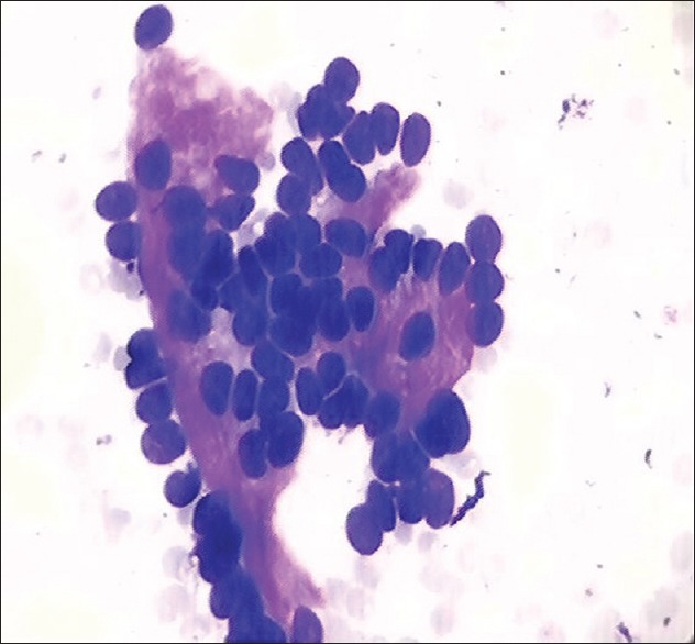
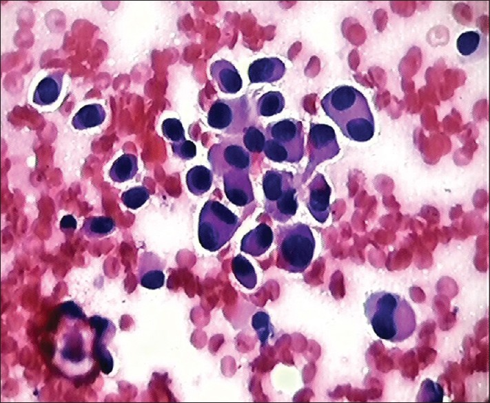
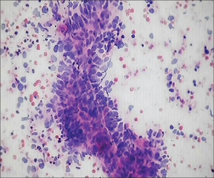
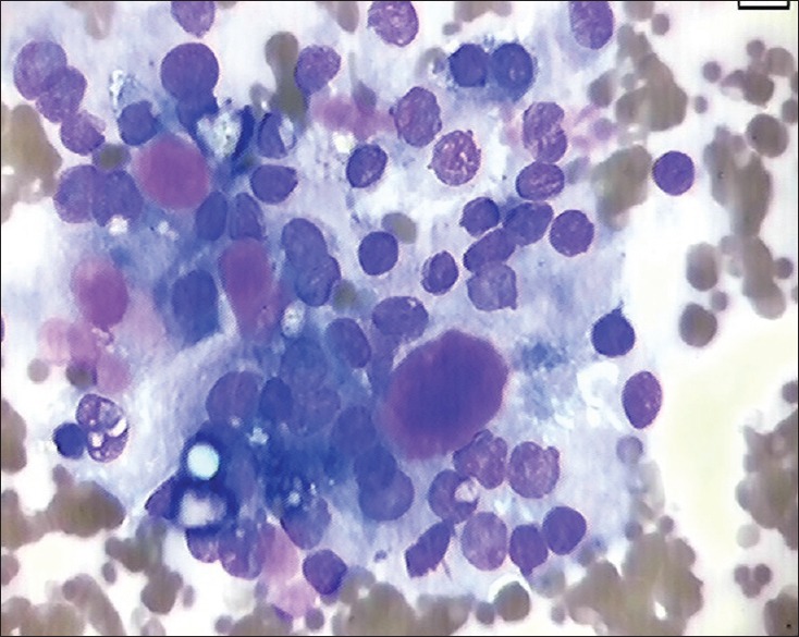
Results: The 170 cytological smears were categorized into two groups: Group 1 adequate aspirations (88.2%), Group 2 inadequate aspirations (11.7%). The adequate aspirations were subdivided as neoplastic (53.33%) and nonneoplastic (46.66%). The distribution of the various neoplastic lesions (80; 53.33%) were 66 (82.5%) benign, 12 (15%) were malignant and 2 (2.5%) were suspicious of malignancy. Among benign neoplasms, the pleomorphic adenoma (62; 93.3%) was the most frequent followed by Warthins tumor (4; 6%). The most common malignant neoplasms were adenoid cystic carcinoma (6; 50%), followed by mucoepidermoid carcinoma (4; 33.3%), malignant lymphoma (1; 8.3%) and metastatic carcinomatous deposits (1; 8.3%). In two cases, cytological picture indicated suspicion for malignancy however specific tumor typing could not be done. The neoplasms occurred more frequently in the parotid gland (65%), followed by submandibular gland (21.3%) and minor salivary glands (13.8%). The nonneoplastic lesions (70) included 68.6% cases of chronic sialadenitis, 17.1% cases were reported as mucocele, 11.4% cases of acute sialadenitis 2.9% cases as tubercular granulomas.
Conclusion: FNA cytology provides useful information on the management of salivary gland lesions and prevents unnecessary surgery in cases of nonneoplastic lesions and identification of malignancy helps the surgeon in deciding type and extent of surgery.
Keywords: Fine-needle aspiration cytology; salivary gland lesions; salivary gland neoplasms.
Conflict of interest statement
There are no conflicts of interest.
Figures

References
-
- Eveson JW, Cawson RA. Salivary gland tumours. A review of 2410 cases with particular reference to histological types, site, age and sex distribution. J Pathol. 1985;146:51–8. - PubMed
-
- Das DK, Petkar MA, Al-Mane NM, Sheikh ZA, Mallik MK, Anim JT. Role of fine needle aspiration cytology in the diagnosis of swellings in the salivary gland regions: A study of 712 cases. Med Princ Pract. 2004;13:95–106. - PubMed
-
- Nguansangiam S, Jesdapatarakul S, Dhanarak N, Sosrisakorn K. Accuracy of fine needle aspiration cytology of salivary gland lesions: Routine diagnostic experience in Bangkok, Thailand. Asian Pac J Cancer Prev. 2012;13:1583–8. - PubMed
-
- Singh Nanda KD, Mehta A, Nanda J. Fine-needle aspiration cytology: A reliable tool in the diagnosis of salivary gland lesions. J Oral Pathol Med. 2012;41:106–12. - PubMed
-
- Al-Khateeb TH, Ababneh KT. Salivary tumors in North Jordanians: A descriptive study. Oral Surg Oral Med Oral Pathol Oral Radiol Endod. 2007;103:e53–9. - PubMed
LinkOut - more resources
Full Text Sources
Other Literature Sources
