

Adenoid ameloblastoma with dentinoid: A rare hybrid variant
- PMID: 28932051
- PMCID: PMC5596692
- DOI: 10.4103/jomfp.JOMFP_53_15
Adenoid ameloblastoma with dentinoid: A rare hybrid variant
Abstract
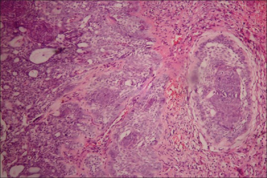
Odontogenic tumors comprise an unusual group of lesions of the jaw and present diverse histological patterns. Derived from the primordial tooth-forming tissues, they represent a heterogeneous group of lesions that range from hamartomas to benign and malignant neoplasms of variable aggressiveness. Sporadic case reports and diverse complex histogenetic source also defy categorization of odontogenic tumors. Many can be diagnosed accurately based on the distinctive clinical, radiological and histopathological presentation. Considerable variations in the clinicopathological presentation of odontogenic tumors can be confusing, increasing the chance of misdiagnosis. An interesting case of adenoid ameloblastoma reported in a 55-year-old male patient in the mandible, presenting with a diverse and intriguing histopathology, is discussed here.
Keywords: Adenomatoid odontogenic tumor; ameloblastoma; architecture; hamartoma; odontogenic tumor.
Conflict of interest statement
There are no conflicts of interest.
Figures


Similar articles
-
Adenoid ameloblastoma with dentinoid: A rare hybrid odontogenic tumor.Indian J Pathol Microbiol. 2024 Apr 1;67(2):441-444. doi: 10.4103/ijpm.ijpm_186_22. Epub 2023 Jul 6. Indian J Pathol Microbiol. 2024. PMID: 38391318
-
Adenoid ameloblastoma with dentinoid is molecularly different from ameloblastomas and adenomatoid odontogenic tumors.J Oral Pathol Med. 2021 Nov;50(10):1067-1071. doi: 10.1111/jop.13243. Epub 2021 Sep 29. J Oral Pathol Med. 2021. PMID: 34549835
-
Diagnostic Enigma of Adenoid Ameloblastoma: Literature Review Based Evidence to Consider It as a New Sub Type of Ameloblastoma.Head Neck Pathol. 2022 Jun;16(2):344-352. doi: 10.1007/s12105-021-01358-w. Epub 2021 Jul 19. Head Neck Pathol. 2022. PMID: 34282559 Free PMC article. Review.
-
Adenoid ameloblastoma with dentinoid.J Oral Maxillofac Pathol. 2012 May;16(2):272-6. doi: 10.4103/0973-029X.99088. J Oral Maxillofac Pathol. 2012. PMID: 22923903 Free PMC article.
-
Atypical plexiform ameloblastoma with dentinoid: adenoid ameloblastoma with dentinoid.J Oral Pathol Med. 2001 Apr;30(4):251-4. doi: 10.1034/j.1600-0714.2001.300410.x. J Oral Pathol Med. 2001. PMID: 11302246 Review.
Cited by
-
Demystifying Histologic Conundrum of Adenoid Ameloblastoma: Case Report with Literature Review.Indian J Otolaryngol Head Neck Surg. 2023 Sep;75(3):2432-2437. doi: 10.1007/s12070-023-03534-6. Epub 2023 Apr 8. Indian J Otolaryngol Head Neck Surg. 2023. PMID: 37636784 Free PMC article.
-
Morphological Features of the Spectrum of Ghost Cell Odontogenic Lesions.Head Neck Pathol. 2024 Oct 15;18(1):102. doi: 10.1007/s12105-024-01688-5. Head Neck Pathol. 2024. PMID: 39404952 No abstract available.
-
Histopathological Insight of a Case of Adenoid Ameloblastoma: A Rare Odontogenic Tumor.Case Rep Dent. 2024 Apr 29;2024:8366045. doi: 10.1155/2024/8366045. eCollection 2024. Case Rep Dent. 2024. PMID: 38716224 Free PMC article.
-
Clinicopathological Considerations in the Treatment of Unicystic Adenoid Ameloblastoma in Young Adults: A Clinical Case.Cureus. 2025 Jul 11;17(7):e87753. doi: 10.7759/cureus.87753. eCollection 2025 Jul. Cureus. 2025. PMID: 40799867 Free PMC article.
-
Update on Odontogenic Tumors: Proceedings of the North American Head and Neck Pathology Society.Head Neck Pathol. 2019 Sep;13(3):457-465. doi: 10.1007/s12105-019-01013-5. Epub 2019 Mar 18. Head Neck Pathol. 2019. PMID: 30887391 Free PMC article.
References
-
- Yamazaki M, Maruyama S, Abé T, Babkair H, Fujita H, Takagi R, et al. Hybrid ameloblastoma and adenomatoid odontogenic tumor: Report of a case and review of hybrid variations in the literature. Oral Surg Oral Med Oral Pathol Oral Radiol. 2014;118:e12–8. - PubMed
-
- Date A, Padhye M, Jagtap D. Adenoid ameloblastoma with dentinoid deposits: Report of a rare case. Sci J. 2008:2.
-
- Paikkatt VJ, Sreedharan S, Kannan VP. Unicystic ameloblastoma of the maxilla: A case report. J Indian Soc Pedod Prev Dent. 2007;25:106–10. - PubMed
-
- Allen CM, Neville BW, Hammond HL. Adenomatoid dentinoma. Report of four cases of an unusual odontogenic lesion. Oral Surg Oral Med Oral Pathol Oral Radiol Endod. 1998;86:313–7. - PubMed
Publication types
LinkOut - more resources
Full Text Sources
Other Literature Sources
