Darier disease: A rare genodermatosis
- PMID: 28932054
- PMCID: PMC5596695
- DOI: 10.4103/jomfp.JOMFP_170_16
Darier disease: A rare genodermatosis
Abstract
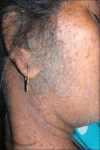
Darier disease (DD), also known as keratosis follicularis or dyskeratosis follicularis, is a rare autosomal dominant genodermatosis with high penetrance and variable expressivity. It is caused by mutations of ATP2A2 gene which encodes the sarco/endoplasmic reticulum Ca2+ ATPase isoform 2. It is clinically manifested by hyperkeratotic papules primarily affecting seborrheic areas on the head, neck and thorax, with less frequent involvement of the oral mucosa. When oral manifestations are present, they primarily affect the palatal and alveolar mucosa, are usually asymptomatic and are discovered in routine dental examination. Histologically, the lesions show suprabasal clefts with acantholytic and dyskeratotic cells. We present a case of 35-year-old female patient with typical clinical and histological features of DD.
Keywords: Autosomal dominant; Darier disease; keratosis follicularis.
Conflict of interest statement
There are no conflicts of interest.
Figures


Similar articles
-
Darier disease: case report with oral manifestations.Med Oral Patol Oral Cir Bucal. 2006 Aug 1;11(5):E404-6. Med Oral Patol Oral Cir Bucal. 2006. PMID: 16878056
-
A Rare Clinical Presentation of Darier's Disease.Case Rep Dermatol Med. 2013;2013:419797. doi: 10.1155/2013/419797. Epub 2013 Mar 20. Case Rep Dermatol Med. 2013. PMID: 23573430 Free PMC article.
-
A Rare Clinical Presentation of Intraoral Darier's Disease.Case Rep Pathol. 2011;2011:181728. doi: 10.1155/2011/181728. Epub 2011 Sep 8. Case Rep Pathol. 2011. PMID: 22937379 Free PMC article.
-
Darier disease.J Dermatol. 2016 Mar;43(3):275-9. doi: 10.1111/1346-8138.13230. J Dermatol. 2016. PMID: 26945535 Review.
-
Darier's disease: epidemiology, pathophysiology, and management.Am J Clin Dermatol. 2003;4(2):97-105. doi: 10.2165/00128071-200304020-00003. Am J Clin Dermatol. 2003. PMID: 12553850 Review.
Cited by
-
Magnesium chloride-calcium carbonate treatment in a pregnant patient with severe Darier disease.JAAD Case Rep. 2023 Nov 30;43:90-92. doi: 10.1016/j.jdcr.2023.11.016. eCollection 2024 Jan. JAAD Case Rep. 2023. PMID: 38223365 Free PMC article. No abstract available.
-
Scratching the Surface: A Comprehensive Guide to Understanding and Managing Vulvovaginal Itching.Am J Clin Dermatol. 2025 May;26(3):361-378. doi: 10.1007/s40257-025-00939-7. Epub 2025 Mar 25. Am J Clin Dermatol. 2025. PMID: 40131720 Free PMC article. Review.
-
Darier's Disease: Report of a Case with Facial Involvement.Case Rep Dermatol. 2019 Dec 10;11(3):327-333. doi: 10.1159/000504925. eCollection 2019 Sep-Dec. Case Rep Dermatol. 2019. PMID: 31911771 Free PMC article.
-
Darier Disease - A Multi-organ Condition?Acta Derm Venereol. 2021 Apr 15;101(4):adv00430. doi: 10.2340/00015555-3770. Acta Derm Venereol. 2021. PMID: 33606037 Free PMC article. Review.
-
Darier's Disease Localized to Face - A Hitherto Unrecognized Presentation.Indian J Dermatol. 2023 Jan-Feb;68(1):110-112. doi: 10.4103/ijd.ijd_448_21. Indian J Dermatol. 2023. PMID: 37151236 Free PMC article. No abstract available.
References
-
- Darier J. De la psorospermose folliculaire végétante. Ann Dermatol Syphiligr. 1889;10:597–612.
-
- White J. A case of keratosis (ichthyosis) follicularis. J Cutan Genitourin Dis. 1889;7:210–9.
-
- Prindiville DE, Stern D. Oral manifestations of Darier's disease. J Oral Surg. 1976;34:1001–6. - PubMed
-
- Munro CS. The phenotype of Darier's disease: Penetrance and expressivity in adults and children. Br J Dermatol. 1992;127:126–30. - PubMed
Publication types
LinkOut - more resources
Full Text Sources
Other Literature Sources
Miscellaneous

