Follicular Hybrid Cyst with Rare Juxtaposition of Epidermal Cyst and Steatocystoma
- PMID: 28932064
- PMCID: PMC5596647
- DOI: 10.4103/ijt.ijt_21_17
Follicular Hybrid Cyst with Rare Juxtaposition of Epidermal Cyst and Steatocystoma
Abstract
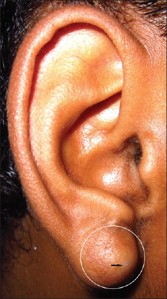
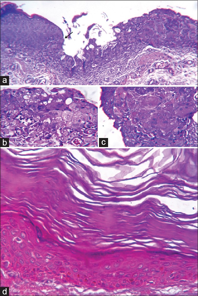
Any cutaneous cyst differentiating toward two or more pilosebaceous components is known as follicular hybrid cyst (FHC). A combination of epidermal and trichilemmal cyst is its most frequent example. Other combinations of pilosebaceous derivatives occur uncommonly as well. The histogenesis of this condition has been controversial. In this latest report, we describe an unusual FHC from the earlobe of a 19-year-old male, which expressed the cohabitation of epidermal cyst and steatocystoma. A sharp transition was noted between the two kinds of epithelial components.
Keywords: Earlobe; epidermal cyst; follicular hybrid cyst; pilosebaceous follicle; steatocystoma.
Conflict of interest statement
There are no conflicts of interest.
Figures
References
-
- Weedon D. Cysts, sinuses and pits. In: Davie B, editor. Weedon's Skin Pathology. 3rd ed. London: Churchill Livingstone, Elsevier; 2010. pp. 442–57.
-
- Lee KY, Kwon YS, Roh MR, Chung KY. A case of a follicular hybrid cyst. Ann Dermatol. 2007;19:153–6.
-
- McGavran MH, Binnington B. Keratinous cysts of the skin. Identification and differentiation of pilar cysts from epidermal cysts. Arch Dermatol. 1966;94:499–508. - PubMed
-
- Kim MS, Lee JH, Son SJ. Hybrid cysts: A clinicopathological study of seven cases. Australas J Dermatol. 2012;53:49–51. - PubMed
Publication types
LinkOut - more resources
Full Text Sources
Other Literature Sources