. 2017 Sep 6:8:673.
doi: 10.3389/fphys.2017.00673. eCollection 2017.

Is Increased Intracellular Calcium in Red Blood Cells a Common Component in the Molecular Mechanism Causing Anemia?

Affiliations

Is Increased Intracellular Calcium in Red Blood Cells a Common Component in the Molecular Mechanism Causing Anemia?

Laura Hertz et al. Front Physiol. .

Abstract

For many hereditary disorders, although the underlying genetic mutation may be known, the molecular mechanism leading to hemolytic anemia is still unclear and needs further investigation. Previous studies revealed an increased intracellular Ca2+ in red blood cells (RBCs) from patients with sickle cell disease, thalassemia, or Gardos channelopathy. Therefore we analyzed RBCs' Ca2+ content from 35 patients with different types of anemia (16 patients with hereditary spherocytosis, 11 patients with hereditary xerocytosis, 5 patients with enzymopathies, and 3 patients with hemolytic anemia of unknown cause). Intracellular Ca2+ in RBCs was measured by fluorescence microscopy using the fluorescent Ca2+ indicator Fluo-4 and subsequent single cell analysis. We found that in RBCs from patients with hereditary spherocytosis and hereditary xerocytosis the intracellular Ca2+ levels were significantly increased compared to healthy control samples. For enzymopathies and hemolytic anemia of unknown cause the intracellular Ca2+ levels in RBCs were not significantly different. These results lead us to the hypothesis that increased Ca2+ levels in RBCs are a shared component in the mechanism causing an accelerated clearance of RBCs from the blood stream in channelopathies such as hereditary xerocytosis and in diseases involving defects of cytoskeletal components like hereditary spherocytosis. Future drug developments should benefit from targeting Ca2+ entry mediating molecular players leading to better therapies for patients.

Keywords: calcium homeostasis; channelopathies; erythrocyte; live cell imaging; rare anemia; spherocytosis; xerocytosis.


Figures
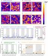
Figure 1
Figure 1
Intracellular Ca2+ content in RBCs from patients with different types of hemolytic anemia. (A–D) Representative fluorescence images of Fluo-4 loaded RBCs from control and patient blood samples. Relative intracellular Ca2+ concentration is scaled from black (lowest) to white (highest). Patients were grouped according to their disease diagnosis: hereditary spherocytosis (blue), hemolytic anemia (purple), enzymopathies (green), hereditary xerocytosis (orange). (E–H) Statistical analysis of the mean Fluo-4 intensity values in arbitrary units from single cells for each group of patients and the corresponding control samples. Controls and patients that belong to the same shipment are highlighted with a white or gray background. Controls are displayed as black boxes, patients are colored in blue (hereditary spherocytosis), purple (unknown hemolytic anemia), green (enzymopathies), and orange (hereditary xerocytosis). Whiskers indicate the range between 10th and 90th percentiles. There is a huge variation in the intensity-correlated Ca2+ concentrations already within the control group of healthy donors. Significance was tested between each patient and the according shipping control sample (always the next control sample on left side) using the Mann–Whitney test (ns denotes p > 0.5, *p ≤ 0.5, ***p ≤ 0.01).
Figure 2
Figure 2
Patient-based statistical analysis of intracellular Ca2+ levels and proposed mechanisms. (A) Statistical analysis of normalized median fluorescence intensity values for all 35 patients (red bar) and 25 healthy shipping controls (black bar). Significance was tested using the Wilcoxon signed-rank test (***p ≤ 0.01). (B) Statistical analysis of normalized median fluorescence intensity values for controls and patients grouped by disease. Number of control samples differs for each disease group. Hereditary spherocytosis (blue): 16 patients and 12 controls, hereditary xerocytosis (orange): 12 patients and 5 controls, hemolytic anemia (purple): 3 patients and 3 controls, enzymopathies (green): 5 patients and 5 controls. Significance was tested using the paired t-test, when data showed a Gaussian distribution (D'Agostino-Pearson normality test), otherwise using the Wilcoxon signed-rank test (ns denotes p > 0.5, ***p ≤ 0.01). (C) Proposed mechanisms leading to increased intracellular Ca2+ levels in diseased RBCs and accordingly to accelerated clearance of cells from the blood stream. Alternative or cumulating Ca2+ entry pathways are highlighted with gray background: increased abundance of NMDA-receptors (NMDAR), e.g., in sickle cell disease, altered activity of Piezo1, e.g., in hereditary xerocytosis, increased activity of Gardos Channel, e.g., in Gardos Channelopathy, or unspecified Ca2+ transport mechanisms. Additionally, ineffective extrusion of Ca2+ due to disruption of ATP pools fueling the plasma membrane Ca2+ ATPase (PMCA) can contribute. Several downstream processes follow Ca2+ overload in RBCs, e.g.: activation of calmodulin by formation of the Ca2+-calmodulin complex (Ca-CaM) and activation of calpain, thereby loosening the cytoskeletal structure; activation of the scramblase (Scr) leading to exposure of phosphatidylserine on the outer leaflet of the membrane; activation of the Gardos channel followed by the efflux of K+, Cl and H2O and consecutive cell shrinkage.

References

    1. Alaarg A., Schiffelers R. M., van Solinge W. W., van Wijk R. (2013). Red blood cell vesiculation in hereditary hemolytic anemia. Front. Physiol. 4:365. 10.3389/fphys.2013.00365 - DOI - PMC - PubMed
    1. Albuisson J., Murthy S. E., Bandell M., Coste B., Louis-dit-Picard H. L. N., Mathur J., et al. . (2013). Dehydrated hereditary stomatocytosis linked to gain-of-function mutations in mechanically activated PIEZO1 ion channels. Nat. Commun. 4, 165–119. 10.1038/ncomms2899 - DOI - PMC - PubMed
    1. Ataga K. I., Reid M., Ballas S. K., Yasin Z., Bigelow C., James L. S., et al. (2011). Improvements in haemolysis and indicators of erythrocyte survival do not correlate with acute vaso-occlusive crises in patients with sickle cell disease: a phase III randomized, placebo-controlled, double-blind study of the Gardos channel blocker senicapoc (ICA-17043). Br. J. Haematol. 153, 92–104. 10.1111/j.1365-2141.2010.08520.x - DOI - PubMed
    1. Bassé F., Stout J. G., Sims P. J., Wiedmer T. (1996). Isolation of an erythrocyte membrane protein that mediates Ca2+-dependent transbilayer movement of phospholipids. J. Biol. Chem. 271, 17205–17210. 10.1074/jbc.271.29.17205 - DOI - PubMed
    1. Bitbol M., Fellmann P., Zachowski A., Devaux P. F. (1987). Ion regulation of phosphatidylserine and phosphatidylethanolamine outside-inside translocation in human erythrocytes. Biochim. Biophys. Acta 904, 268–282. 10.1016/0005-2736(87)90376-2 - DOI - PubMed

LinkOut - more resources