

Incontinentia pigmenti in a child with suspected retinoblastoma
- PMID: 28932485
- PMCID: PMC5603187
- DOI: 10.1186/s40942-017-0088-5
Incontinentia pigmenti in a child with suspected retinoblastoma
Abstract
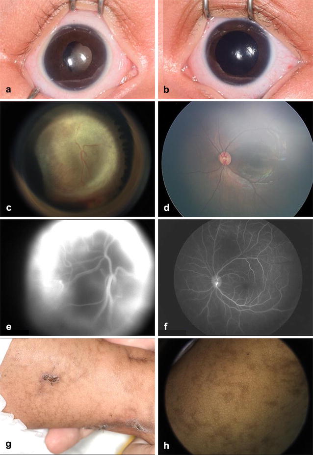
Background: Incontinentia pigmenti is a rare X-linked dominant syndrome caused by mutation in the NEMO/IKKgamma gene, and characterized by a spectrum of cutaneous, ocular, neurologic and dental abnormalities. In the eye, findings include retinal vascular non-perfusion, occasionally with traction retinal detachment, retinal fibrosis, and retinal pigment epithelium defects. These findings can resemble retinoblastoma, especially when vitreoretinal fibrosis produces leukocoria.
Case report: A 2-month-old girl born full-term presented with leukocoria, suspicious for retinoblastoma. She was found to have an ischemic retrolental fibrovascular retinal detachment. In addition, there was linear cutaneous hyperpigmentation, diagnostic of incontinentia pigmenti.
Conclusions: Retinoblastoma can be a challenge to diagnose. There are numerous simulating lesions that can present with leukocoria and retinal detachment, including incontinentia pigmenti. Recognition of the cutaneous features of incontinentia pigmenti contributes to early detection of related ophthalmologic, neurologic and dental abnormalities.
Keywords: Bloch–Sulzberger syndrome; Eye; Incontinentia pigmenti; Pseudoretinoblastoma; Retinal detachment; Retinoblastoma.
Figures
Similar articles
-
Incontinentia pigmenti (Bloch-Sulzberger-syndrome): case report and differential diagnosisto related dermato-ocular syndromes.Ophthalmologica. 1999;213(1):63-9. doi: 10.1159/000027396. Ophthalmologica. 1999. PMID: 9838260
-
Incontinentia pigmenti (Bloch-Sulzberger syndrome): a case report and review of the ocular pathological features.Can J Ophthalmol. 1991 Jun;26(4):229-37. Can J Ophthalmol. 1991. PMID: 1889027
-
Incontinentia pigmenti: a rare cause of retinal vasculitis in children.Tunis Med. 2008 Dec;86(12):1079-81. Tunis Med. 2008. PMID: 19213518
-
Incontinentia pigmenti: a review and update on the molecular basis of pathophysiology.J Am Acad Dermatol. 2002 Aug;47(2):169-87; quiz 188-90. doi: 10.1067/mjd.2002.125949. J Am Acad Dermatol. 2002. PMID: 12140463 Review.
-
Incontinentia Pigmenti.Actas Dermosifiliogr (Engl Ed). 2019 May;110(4):273-278. doi: 10.1016/j.ad.2018.10.004. Epub 2019 Jan 17. Actas Dermosifiliogr (Engl Ed). 2019. PMID: 30660327 Review. English, Spanish.
Cited by
-
Central nervous system anomalies in 41 Chinese children incontinentia pigmenti.BMC Neurosci. 2024 May 21;25(1):25. doi: 10.1186/s12868-024-00872-1. BMC Neurosci. 2024. PMID: 38773385 Free PMC article.
-
Uncovering incontinentia pigmenti: From DNA sequence to pathophysiology.Front Pediatr. 2022 Sep 6;10:900606. doi: 10.3389/fped.2022.900606. eCollection 2022. Front Pediatr. 2022. PMID: 36147820 Free PMC article. Review.
References
Publication types
LinkOut - more resources
Full Text Sources
Other Literature Sources
Miscellaneous
