Review
. 2017 Aug 30;2(8):362-371.
doi: 10.1302/2058-5241.2.160006. eCollection 2017 Aug.

Medial elbow pain

Affiliations
Review

Medial elbow pain

Raul Barco et al. EFORT Open Rev. .

Abstract

Medial elbow pain is uncommon when compared with lateral elbow pain.Medial epicondylitis is an uncommon diagnosis and can be confused with other sources of pain.Overhead throwers and workers lifting heavy objects are at increased risk of medial elbow pain.Differential diagnosis includes ulnar nerve disorders, cervical radiculopathy, injured ulnar collateral ligament, altered distal triceps anatomy or joint disorders.Children with medial elbow pain have to be assessed for 'Little League elbow' and fractures of the medial epicondyle following a traumatic event.This paper is primarily focused on the differential diagnosis of medial elbow pain with basic recommendations on treatment strategies. Cite this article: EFORT Open Rev 2017;2:362-371. DOI: 10.1302/2058-5241.2.160006.

Keywords: elbow; medial elbow pain; medial epicondylitis; sports; ulnar collateral ligament; ulnar neuritis.


Conflict of interest statement

ICMJE Conflict of interest statement: Raul Barco is a board member of SECEC, ESSKA and SECHC, and reports consultancy and payment for lectures from Exactech, Conmed and Acumed, outside the submitted work. Samuel A. Antuña is a board member of the Journal of Shoulder and Elbow Surgery, consults for Exactech and receives payment for development of educational presentations from Zimmer Biomet and Acumed, outside the submitted work.

Figures

Fig. 1
Fig. 1
Patients with medial epicondylitis have pain on resisted flexion and/or pronation. Those patients with more chronic symptoms may exhibit pain with resisted elbow flexion. This manoeuvre, shown in this clinical picture, includes resisted elbow and wrist flexion and resisted pronation, thus, is very sensitive. The patient is asked to take the hand as if to wash their face and the examiner places resistance on the radial border of the hand. Pain on the medial epicondyle is generally reproduced in patients with medial epicondylitis.
Fig. 2
Fig. 2
This picture depicts exploring the elbow with the ‘moving valgus test’ developed by O’Driscoll et al. The patient is seated with the shoulder locked in maximum external rotation. The examiner places the elbow through a range of movement while applying a valgus torque on the elbow throughout the exploration. Patients with medial elbow instability typically have maximum pain on the medial side of the elbow between 75° to 95º of elbow flexion. Pain in terminal extension and pronation may be used to detect valgus overload syndrome.
Fig. 3
Fig. 3
A medial approach at the interval between the flexor carpi radialis and pronator teres is performed with care to protect the medial antebrachial cutaneous nerve branches: a) the fascia is opened and the degenerative tissue is debrided to healthy tissue; b) the bone is slightly decorticated to promote healing and a bony anchor is inserted; c) the remnant tendinous healthy tissue is reinserted to bone with the aid of the bony anchor and the rest of the fascia is closed in a standard manner.
Fig. 4
Fig. 4
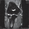
In this T2 fat-saturated coronal MRI view, a partial tear of the medial collateral ligament from the medial epicondyle is observed as a high intensity signal (white arrow).
Fig. 5
Fig. 5
Loose bodies and osteophytes are removed arthroscopically in a patient with chronic medial collateral ligament insufficiency prior to ligament reconstruction. The image corresponds to a posterior viewing portal of a right elbow with a tissue grasper inserted through a posterolateral portal removing a posteromedial loose osteophyte.
Fig. 6
Fig. 6
When the triceps distal insertion extends medially it may predispose to elbow snapping. In this intra-operative image of a left elbow, we observe exposure of the ulnar nerve and release of the medial extension of the triceps which can be removed or flipped on its long axis and reinserted to the native triceps. This will remove the snapping generated by the triceps. The ulnar nerve usually needs an anterior subcutaneous transposition at the end of the procedure to prevent snapping from the ulnar nerve over the medial epicondyle.
Fig. 7
Fig. 7
a) A displaced and comminuted medial epicondyle fracture is observed in an seven-year-old boy. b) Operative fixation of the fracture is performed with a screw. Intra-operative reduction with a Kirschner-wire assists in the reduction and is removed at the end of the procedure.

References

    1. Shiri R, Viikari-Juntura E, Varonen H, Heliövaara M. Prevalence and determinants of lateral and medial epicondylitis: a population study. Am J Epidemiol 2006;164:1065-1074. - PubMed
    1. Shiri R, Viikari-Juntura E. Lateral and medial epicondylitis: role of occupational factors. Best Pract Res Clin Rheumatol 2011;25:43-57. - PubMed
    1. David TS. Medial elbow pain in the throwing athlete. Orthopedics 2003;26:94-105. - PubMed
    1. Lynch JR, Waitayawinyu T, Hanel DP, Trumble TE. Medial collateral ligament injury in the overhand-throwing athlete. J Hand Surg Am 2008;33:430-437. - PubMed
    1. Cesmebasi A, O’Driscoll SW, Smith J, Skinner JA, Spinner RJ. The snapping medial antebrachial cutaneous nerve. Clin Anat 2015;28:872-877. - PubMed

LinkOut - more resources