

Predictors of conversion to thoracotomy during video-assisted thoracoscopic surgery lobectomy in lung cancer: additional predictive value of FDG-PET/CT in a tuberculosis endemic region
- PMID: 28932548
- PMCID: PMC5594172
- DOI: 10.21037/jtd.2017.07.40
Predictors of conversion to thoracotomy during video-assisted thoracoscopic surgery lobectomy in lung cancer: additional predictive value of FDG-PET/CT in a tuberculosis endemic region
Abstract
Background: To evaluate the added clinical value of 18F-fluorodeoxyglucose (FDG) positron emission tomography/computed tomography (PET/CT) scans to chest CT imaging in predicting the conversion to thoracotomy during video-assisted thoracoscopic surgery (VATS) lobectomy in patients with lung cancer.
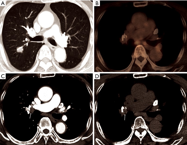
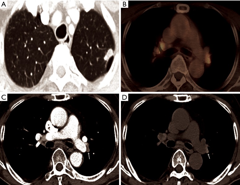
Methods: This is a retrospective study of 235 consecutive patients who underwent planned VATS lobectomy for primary lung cancer between 2011 and 2015. CT images were interpreted in terms of the presence and the attenuation of peribronchial lymph nodes (PLN) and peribronchial cuffs of soft (PCS) tissue, pleural calcification, and parenchymal calcified nodule. On FDG PET/CT images, anthracofibrotic lymph node was considered present when high FDG uptake (SUVmax >3.5) was observed on PET/CT images corresponded to PLN or PCS on chest CT.
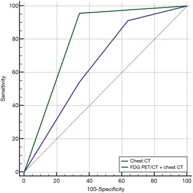
Results: Among the 235 patients undergoing attempted VATS lobectomy, 55 (23.4%) underwent conversion to thoracotomy. Multivariate logistic regression analysis revealed that the attenuation of PLN or PCS on chest CT (OR, 2.57; 95% CI, 1.328-4.380, 0.005) was an only independent predictor of conversion. The ROC curve showed that combined FDG PET/CT and chest CT reading [areas under curve (AUC), 0.847 (95% CI, 0.795-0.891)] was significantly better than that of chest CT scans alone [AUC, 0.655 (95% CI, 0.50-0.751)] in predicting conversion (P=0.024).
Conclusions: The addition of FDG PET/CT scanning to chest CT imaging provides better performance for predicting conversion to thoracotomy during VATS lobectomy in lung cancer patients. Therefore, in lung cancer patients undergoing surgical resection, FDG PET/CT can provide additional reliable information in selecting the appropriate surgical approach for a lobectomy.
Keywords: Lung neoplasms; peribronchial lymph node (PLN); positron-emission tomography; thoracotomy; video-assisted thoracic surgery (VATS).
Conflict of interest statement
Conflicts of Interest: The authors have no conflicts of interest to declare.
Figures
References
-
- Nomori H, Ohtsuka T, Horio H, et al. Difference in the impairment of vital capacity and 6-minute walking after a lobectomy performed by thoracoscopic surgery, an anterior limited thoracotomy, an anteroaxillary thoracotomy, and a posterolateral thoracotomy. Surg Today 2003;33:7-12. 10.1007/s005950300001 - DOI - PubMed
LinkOut - more resources
Full Text Sources
Other Literature Sources