Review
. 2017 Aug;9(8):2619-2639.
doi: 10.21037/jtd.2017.07.68.

The technique of endoscopic airway tumor treatment

Affiliations
Review

The technique of endoscopic airway tumor treatment

Simone Scarlata et al. J Thorac Dis. 2017 Aug.

Abstract

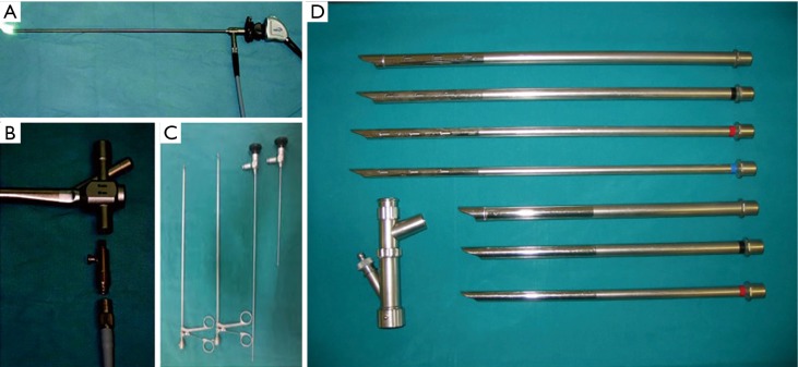
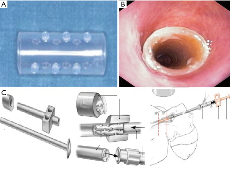
More than half of primary lung cancers are not resectable at diagnosis and 40% of deaths may be secondary to loco-regional disease. Many of these patients suffer from symptoms related to airways obstruction. Indications for therapeutic endoscopic treatment are palliation of dyspnea and other obstructive symptoms in advanced cancerous lesions and cure of early lung cancer. Bronchoscopic management is also indicated for all those patients suffering from benign or minimally invasive neoplasm who are not suitable for surgery due to their clinical conditions. Clinicians should select cases, evaluating tumor features (size, location) and patient characteristics (age, lung function impairment) to choose the most appropriate endoscopic technique. Laser therapy, electrocautery, cryotherapy and stenting are well-described techniques for the palliation of symptoms due to airway involvement and local treatment of endobronchial lesions. Newer technologies, with an established role in clinical practice, are endobronchial ultrasound (EBUS), autofluorescence bronchoscopy (AFB), and narrow band imaging (NBI). Other techniques, such as endobronchial intra-tumoral chemotherapy (EITC), EBUS-guided-transbronchial needle injection or bronchoscopy-guided radiofrequency ablation (RFA), are in development for the use within the airways. These endobronchial interventions are important adjuncts in the multimodality management of lung cancer and should become standard considerations in the management of patients with advanced lung cancer, benign or otherwise not approachable central airway lesions. We aimed at revising several endobronchial treatment modalities that can augment standard antitumor therapies for advanced lung cancer, including rigid and flexible bronchoscopy, laser therapy, endobronchial prosthesis, and photodynamic therapy (PDT).

Keywords: Interventional pulmonology; lung cancer; tracheo-bronchial malignancies; tracheobronchial endoscopy; treatment.


Conflict of interest statement

Conflicts of Interest: The authors have no conflicts of interest to declare.

Figures

Figure 1
Figure 1
Bronchoscopic view of a benign tumor (lipoma) of the anterior tracheal wall.
Figure 2
Figure 2
Endoscopic treatment of early stage trachea-bronchial tumors. (A) In situ carcinoma of the right side tracheal wall; (B) laser resection of the lesion.
Figure 3
Figure 3
Broad based carcinoid of the main left bronchus. Note the smooth, vascular, surface and the soft masses covered with intact bronchial epithelium.
Figure 4
Figure 4
Endoscopic treatment of carcinoid tumors. (A) Three steps laser eradication of a centrally located, totally endobronchial, small based carcinoid tumor of the distal main left bronchus; (B) excision of the tumor by laser resection of the base implant; (C) 30–60 days delayed endoscopic re-evaluation and laser treatment of the implant scar; (D) middle and long term endoscopic control for excluding recurrence.
Figure 5
Figure 5
Bronchoscopic view of a malignant endobronchial obstruction.
Figure 6
Figure 6
The intensity of the autofluorescence differs substantially between normal and tumorous tissues, which allows visualization of cancers and precancerous lesions in bronchi. According to the different systems, normal bronchial mucosa appears green or gray, while cancers and pre-invasive lesions appear brown, or brown-red and dark. (A) White light videobronchoscopy revealing a small bronchial nodule at the bifurcation of the right anterior segmental bronchus and lateral segmental bronchus; (B) under auto-fluorescence imaging, the nodule showed reddish brown area with defined margin on normal mucosa appearing green color.
Figure 7
Figure 7
A narrow banding filter cuts all wavelengths in illumination except for narrow bands in the blue and green spectrum and allows visualize the microvascular structure on mucosal surfaces.
Figure 8
Figure 8
Standard bronchoscopic suite design.
Figure 9
Figure 9
Rigid bronchoscopy equipment.
Figure 10
Figure 10
Laser assisted endoscopic resection of a benign endobronchial neoplasm (83). Available online: http://www.asvide.com/articles/1642
Figure 11
Figure 11
Airway stenting. (A) Silicone stent features, and (B) after placement; (C) charging device and releasing technique.

Similar articles

Cited by

References

    1. Cancer Research UK. Lung cancer incidence statistics. Date last accessed: January 3, 2015. Date last updated: May 29, 2014. Available online: www.cancerresearchuk.org/cancer-info/cancerstats/types/lung/incidence/uk...
    1. Ernst A, Feller-Kopman D, Becker HD, et al. Central airway obstruction. Am J Respir Crit Care Med. 2004;169:1278-97. 10.1164/rccm.200210-1181SO - DOI - PubMed
    1. Wilson RW, Kirejezyk W. Pathological and radiological correlation of endobronchial neoplasm. Part I. Benign tumors. Ann Diagn Pathol 1997;1:31-46. 10.1016/S1092-9134(97)80007-X - DOI - PubMed
    1. Scarlata S, Graziano P, Lucantoni G, et al. Endoscopic treatment of primary benign central airway tumors: Results from a large consecutive case series and decision making flow chart to address bronchoscopic excision. Eur J Surg Oncol. 2015;41:1437-42. 10.1016/j.ejso.2015.08.157 - DOI - PubMed
    1. Kurimoto N, Murayama M, Yoshioka S, et al. Assessment of usefulness of endobronchial ultrasonography in determination of depth of tracheobronchial tumor invasion. Chest 1999;115:1500-6. 10.1378/chest.115.6.1500 - DOI - PubMed

LinkOut - more resources