. 2017 Jun 16;6(9):e1339853.
doi: 10.1080/2162402X.2017.1339853. eCollection 2017.

The anti-SLAMF7 antibody elotuzumab mediates NK cell activation through both CD16-dependent and -independent mechanisms

Affiliations

The anti-SLAMF7 antibody elotuzumab mediates NK cell activation through both CD16-dependent and -independent mechanisms

Tatiana Pazina et al. Oncoimmunology. .

Abstract

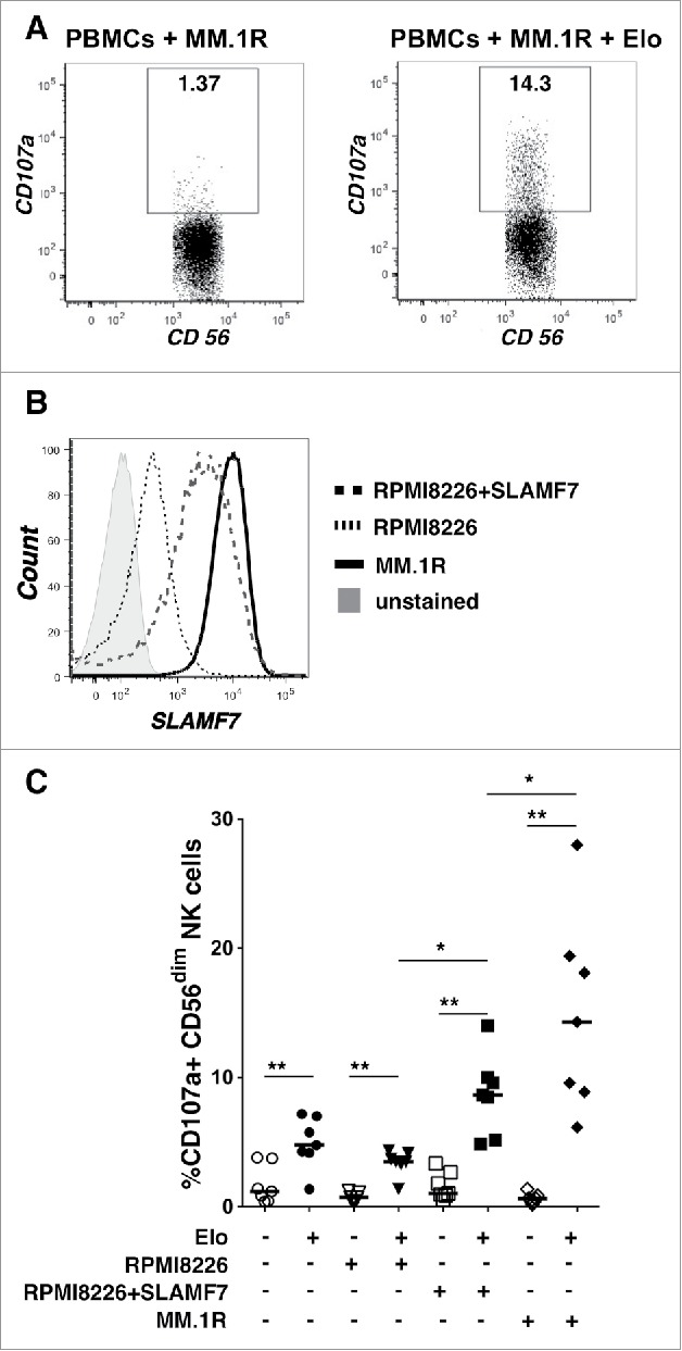
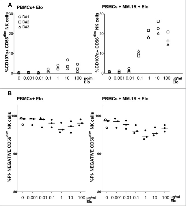
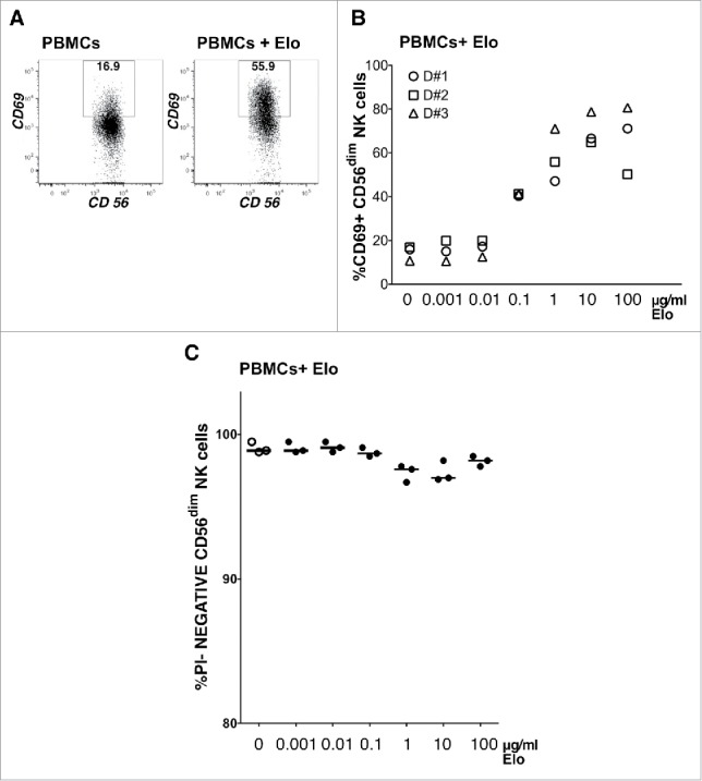
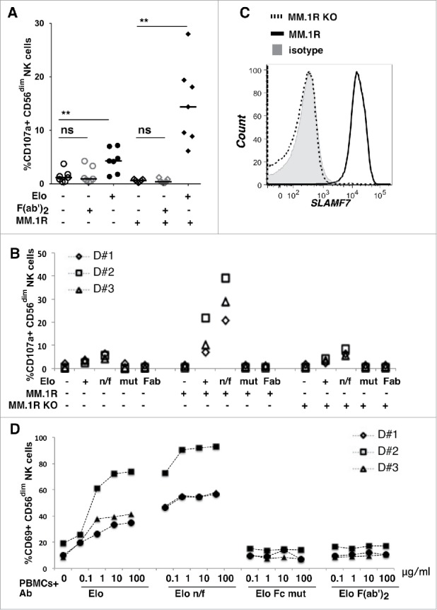
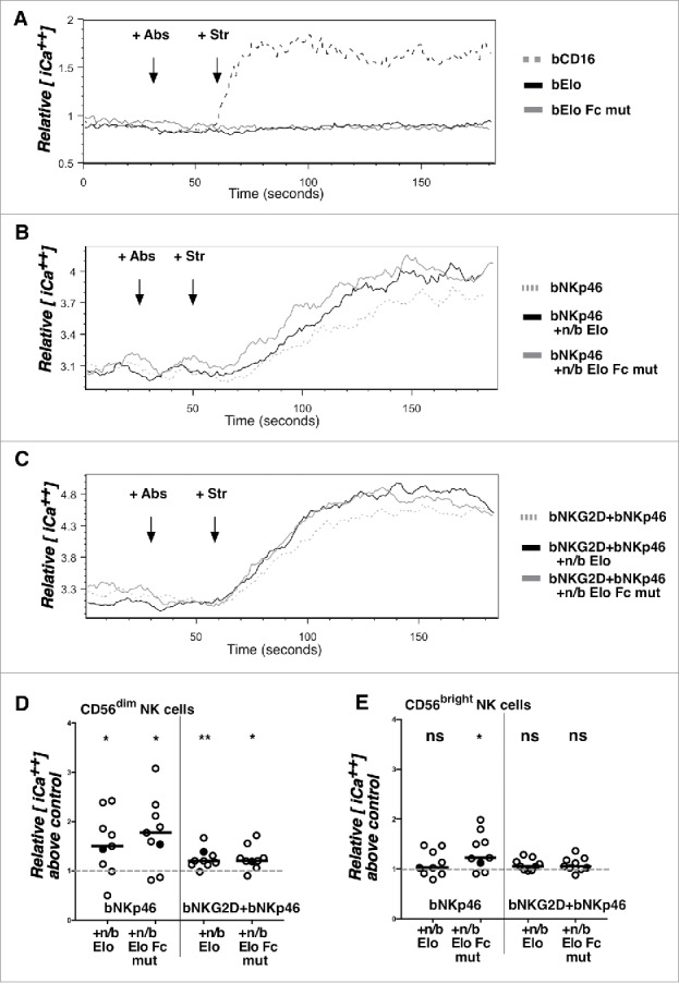
Elotuzumab is a humanized therapeutic monoclonal antibody directed to the surface glycoprotein SLAMF7 (CS1, CRACC, CD319), which is highly expressed on multiple myeloma (MM) tumor cells. Improved clinical outcomes have been observed following treatment of MM patients with elotuzumab in combination with lenalidomide or bortezomib. Previous work showed that elotuzumab stimulates NK cell-mediated antibody-dependent cellular cytotoxicity (ADCC), via Fc-domain engagement with FcγRIIIa (CD16). SLAMF7 is also expressed on NK cells, where it can transmit stimulatory signals. We tested whether elotuzumab can directly activate NK cells via ligation with SLAMF7 on NK cells in addition to targeting ADCC through CD16. We show that elotuzumab strongly promoted degranulation and activation of NK cells in a CD16-dependent manner, and a non-fucosylated form of elotuzumab with higher affinity to CD16 exhibited enhanced potency. Using F(ab')2 or Fc-mutant forms of the antibody, the direct binding of elotuzumab to SLAMF7 alone could not stimulate measurable CD69 expression or degranulation of NK cells. However, the addition of soluble elotuzumab could costimulate calcium signaling responses triggered by multimeric engagement of NKp46 and NKG2D in a CD16-independent manner. Thus, while elotuzumab primarily stimulates NK cells through CD16, it can also transduce effective "trans"-costimulatory signals upon direct engagement with SLAMF7, since these responses did not require direct co-engagement with the activating receptors. Trans-costimulation by elotuzumab has potential to reduce activation thresholds of other NK cell receptors engaging with their ligands on myeloma target cell surfaces, thereby potentially further increasing NK cell responsiveness in patients.

Keywords: ADCC; NK cells; SLAMF7; elotuzumab; multiple myeloma.


Figures

Figure 1.
Figure 1.
Elotuzumab promotes NK cell degranulation that correlates with SLAMF7 expression on myeloma target cell lines. A) NK cell degranulation (CD107a+) from a representative healthy donor after 2 hours incubation with MM.1R targets alone (left; PBMC to target ratio 1:1) or with 1µg/ml Elo. Percentage degranulating CD56dim NK cells is indicated in the box gates. B) SLAMF7 expression on myeloma target cell lines using biotinylated Elo and streptavidin-APC. Unstained cells = gray shaded, parental RPMI8226 = dotted (MFI 422), SLAMF7-transduced RPMI8226 = dashed (MFI 2254), and MM.1R = solid (MFI 10,973). C) Degranulation responses by NK cells in PBMCs from healthy donors (n = 7) alone (circles) or exposed to RPMI-8226 (inverted triangles), RPMI-8226+SLAMF7 (squares) or MM.1R target cells (diamonds) in the presence (+; filled) or absence (-; empty) of 1µg/ml Elo. Horizontal lines = medians; each datapoint = a healthy donor. Overhead bars mark statistical comparisons between indicated groups using one-sided or 2-sided Wilcoxon matched-pairs signed rank test, **- P < 0.01, *-P < 0.05.
Figure 2.
Figure 2.
NK cell degranulation correlates with elotuzumab concentration. A) Percentage CD56dim NK cells in PBMC from 3 healthy donors degranulating after 2 hours with various concentrations (0.001–100µg/ml) of Elo alone (left) or with MM.1R targets (right; PBMC to target cell ratio 1:1). B) Viability of CD56dim NK cells as the percentage of propidium iodide (PI)-negative CD56dim NK cells for each condition.
Figure 3.
Figure 3.
NK cell activation correlates with elotuzumab concentration. A) CD56dim NK cell activation from a representative healthy donor was measured as percentage of CD69+ cells after overnight incubation without (left) or with 1ug/ml Elo (right), as indicated in the box gate. B) Percentage of CD56dim NK cells in PBMCs from 3 healthy donors expressing CD69 after overnight exposure to indicated concentrations of Elo alone. C) Viability of CD56dim NK cells as the percentage of PI-negative CD56dim NK cells for each condition.
Figure 4.
Figure 4.
NK cell degranulation and activation requires the Fc portion of elotuzumab. A) Percentage of CD56dim NK cells that degranulated (CD107a+) in PBMCs from 7 healthy donors after exposure to whole Elo or F(ab')2 Elo at 1ug/ml with (+) or without (-) MM.1R targets. Horizontal lines are medians. Overhead bars mark statistical comparisons between indicated groups using one-sided Wilcoxon matched-pairs signed rank test, **- P < 0.01, ns – not significant. B) NK cell degranulation in PBMCs from 3 healthy donors after exposure to various forms of Elo alone (+ = native mAb, n/f = non-fucosylated Elo, mut = Elo Fc mutant, and Fab = F(ab')2 Elo; 1ug/ml each) with (+) or without (-) MM.1R or SLAMF7-deficient (KO) MM.1R targets (PBMC to target ratio 1:1). C) SLAMF7 expression on myeloma target cells using biotinylated Elo and streptavidin-APC. Isotype treated = gray shaded histogram, SLAMF7-deficient (KO) MM.1R = dashed line, and MM.1R = solid line. D) Activation of CD56dim NK cells in PBMC from 3 healthy donors upon exposure to indicated concentrations (µg/ml) of various forms of Elo overnight as a percentage CD56dim NK cells expressing CD69.
Figure 5.
Figure 5.
Co-activation of calcium signaling responses by SLAMF7 engagement with elotuzumab. A) PBMCs from healthy donors were loaded with indo-1-AM and stained for CD56 and CD3 before analysis by flow cytometry as in Materials and Methods. Cells were analyzed for 30 seconds, treated with antibodies (+Abs) [biotinylated anti-CD16 (bCD16; dashed gray line), bElo (solid black line), or bElo Fc mutant (solid gray line)], and analyzed for 30 seconds, exposed to streptavidin (+Str) to aggregate antibodies, and analyzed for 2 additional minutes. Relative mean intracellular calcium concentration [iCa2+] is indicated from the mean ratio of violet (405 nm) to blue (485 nm) emission over time in gated CD56dim NK cells. B) PBMCs were stimulated using biotinylated anti-NKp46 alone (+bNKp46; dashed gray line) or in combination with non-biotinylated (+n/b) Elo (black solid line) or +n/b Elo Fc mutant (gray solid line) at 30 seconds, then streptavidin 30 seconds later. C) PBMCs were stimulated using biotinylated anti-NKG2D + biotinylated anti-NKp46 mAbs (bNKG2D+bNKp46; dashed gray line) or these 2 biotinylated mAbs in combination with non-biotinylated (+n/b) Elo (black solid line) or +n/b Elo Fc mutant (gray solid line) at 30 seconds, then streptavidin 30 seconds later. D,E) Individual icons represent the increase of relative mean intracellular calcium concentration above control (+bNKp46 or bNKG2D+bNKp46 alone normalized to a value of 1) for CD56dim NK cells (D) or CD56bright NK cells (E) from 9 healthy donors. Relative intracellular calcium values for the gated NK cell populations from each donor were calculated by determining the area under the curve for mean 405nm/485nm emission ratios between 120–180 seconds in Elo-treated samples (+bNKp46 ± bNKG2D + Str as indicated) divided by area under the curve values for the same time window in samples treated with bNKp46 + Str alone (left) or bNKp46 + bNKG2D + Str alone (right). Conditions with and without n/b Elo or Elo Fc mutant were compared by 2-sided Wilcoxon matched-pairs signed rank test, *- P < 0.05, **- P < 0.01. Filled circles indicate ratio values for the representative donor shown in panels (B) and C.

References

    1. Palumbo A, Anderson K. Multiple myeloma. N Eng J Med 2011; 364:1046-60; PMID:21410373; https://doi.org/10.1056/NEJMra1011442 - DOI - PubMed
    1. Siegel RL, Miller KD, Jemal A. Cancer statistics, 2016. CA Cancer J Clin 2016; 66:7-30 - PubMed
    1. Mahindra A, Laubach J, Raje N, Munshi N, Richardson PG, Anderson K. Latest advances and current challenges in the treatment of multiple myeloma. Nat Rev Clin Oncol 2012; 9:135-43; PMID:22349016; https://doi.org/10.1038/nrclinonc.2012.15 - DOI - PubMed
    1. Weiner LM, Surana R, Wang S. Monoclonal antibodies: versatile platforms for cancer immunotherapy. Nat Rev Immunol 2010; 10:317-27; PMID:20414205; https://doi.org/10.1038/nri2744 - DOI - PMC - PubMed
    1. Palumbo A, Sonneveld P. Preclinical and clinical evaluation of elotuzumab, a SLAMF7-targeted humanized monoclonal antibody in development for multiple myeloma. Expert Rev Hematol 2015; 8(4):481-91:1-11 - PubMed

Publication types