. 2017 Jun 20;6(9):e1341031.
doi: 10.1080/2162402X.2017.1341031. eCollection 2017.

Romidepsin alone or in combination with anti-CD20 chimeric antigen receptor expanded natural killer cells targeting Burkitt lymphoma in vitro and in immunodeficient mice

Affiliations

Romidepsin alone or in combination with anti-CD20 chimeric antigen receptor expanded natural killer cells targeting Burkitt lymphoma in vitro and in immunodeficient mice

Yaya Chu et al. Oncoimmunology. .

Abstract

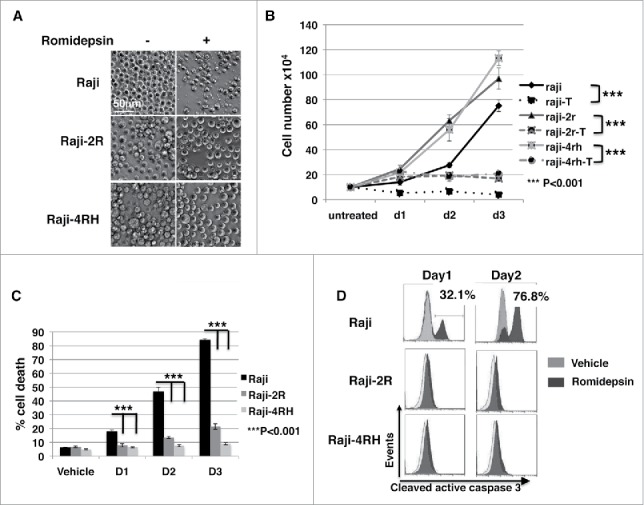
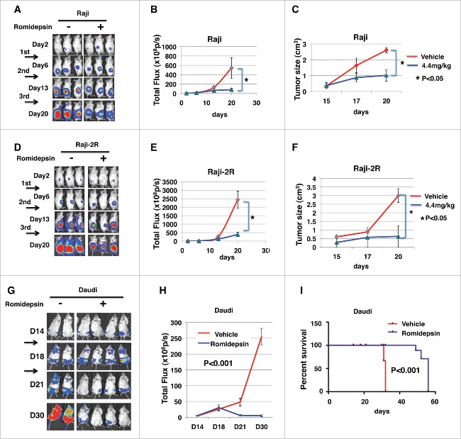
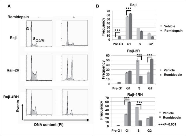
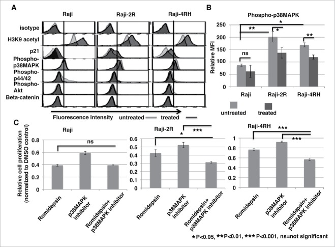
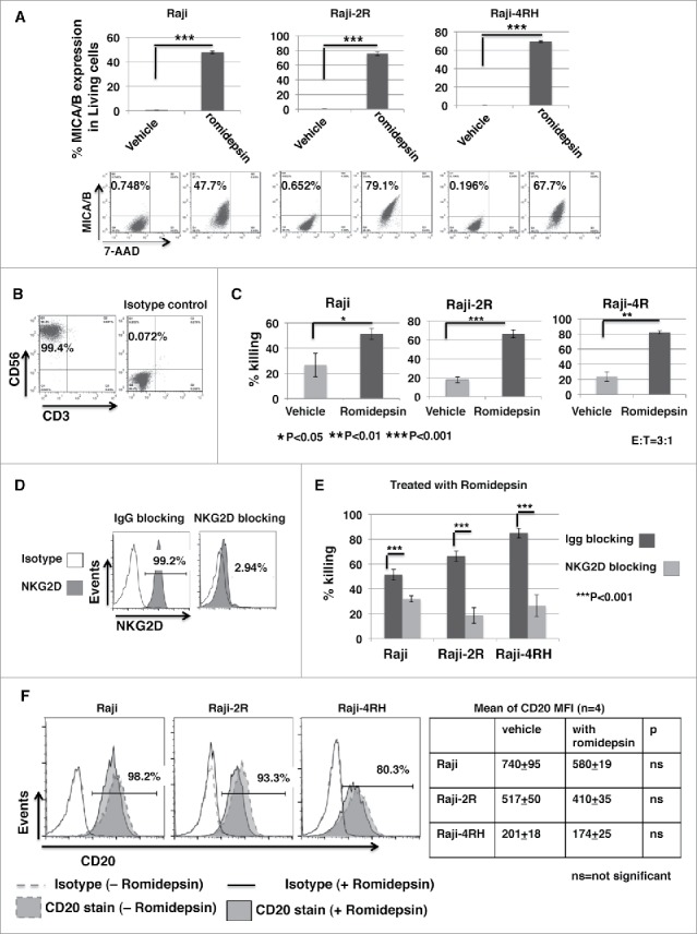
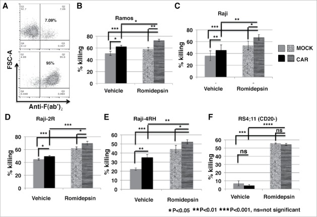
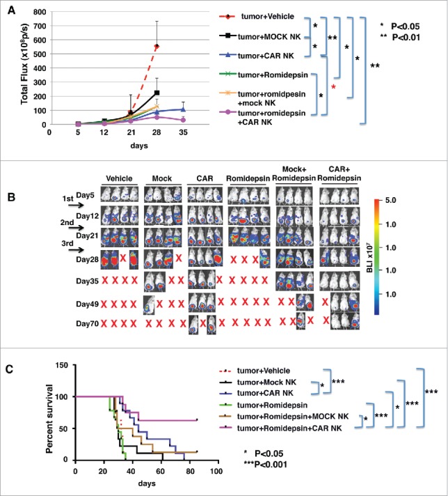
Facilitating the development of alternative targeted therapeutic strategies is urgently required to improve outcome or circumvent chemotherapy resistance in children, adolescents, and adults with recurrent/refractory de novo mature B-cell (CD20) non-Hodgkin lymphoma, including Burkitt lymphoma (BL). Romidepsin, a histone deacetylase inhibitor (HDACi), has been used to treat cutaneous T-cell lymphoma. We have demonstrated the significant anti-tumor effect of anti-CD20 chimeric antigen receptor (CAR) modified expanded peripheral blood natural killer (exPBNK) against rituximab-sensitive and -resistant BL. This study examined the anti-tumor activity of romidepsin alone and in combination with anti-CD20 CAR exPBNKs against rituximab-sensitive and -resistant BL in vitro and in vivo. We found that romidepsin significantly inhibited both rituximab-sensitive and -resistant BL cell proliferation in vitro (P < 0.001) and induced cell death in rituximab-sensitive Raji (P < 0.001) and cell cycle arrest in rituximab-resistant Raji-2R and Raji-4RH (P < 0.001). Consistent with in vitro observations, we also found romidepsin significantly inhibited the growth of rituximab-sensitive and -resistant BL in BL xenografted NSG mice. We also demonstrated that romidpesin significantly induced the expression of Natural Killer Group 2, Member D (NKG2D) ligands MICA/B in both rituximab-sensitive and -resistant BL cells (P < 0.001) resulting in enhancement of exPBNK in vitro cytotoxicity through NKG2D. Finally, we observed the combination of romidepsin and anti-CD20 CAR exPBNK significantly induced cell death in BL cells in vitro, reduced tumor burden and enhanced survival in humanized BL xenografted NSG mice (p < 0.05). Our data suggests that romidepsin is an active HDAC inhibitor that also potentiates expanded NK and anti-CD20 CAR exPBNK activity against rituximab-sensitive and -resistant BL.

Keywords: anti-CD20 chimeric antigen receptor; expanded Natural Killer Cells; rituximab sensitive and resistant Burkitt Lymphoma; romidepsin; targeted immunotherapy.


Figures

Figure 1.
Figure 1.
Romidepsin significantly inhibits cell proliferation in both rituximab sensitive and resistant cells and stimulates cell death in rituximab sensitive cells. CD20+ rituximab sensitive Raji and resistant Raji-2R and Raji-4RH cells were treated with or without 10ng/ml romidepsin for 3 days. A, cell phenotypic changes under light microscopy (Carl Zeiss, Thornwood, NY)) are shown at day 3 (original magnification 200x). B, cell proliferation curves were generated with trypan blue staining of living cells and counting living cells with hemocytometer. C, the percentage of the dead cells was gated with 7-AAD+ by flow cytometry analysis. Average values are reported as the mean ± SEM. P values using unpaired student t test were noted in B and C respectively. D, intracellular caspase 3 activation was monitored by flow cytometry analysis at day 1 and day 2. DMSO was added in equal amounts and served as a vehicle control.
Figure 2.
Figure 2.
Romidepsin significantly inhibits Raji and Raji-2R cells growth in xenografted mice. 5 × 10⁵ of Raji-Luc or Raji-2R-Luc cells were s.c. injected in the right flanks of NSG mice. 3 d after tumor inoculation, mice were randomized to equalize tumor burden and injected i.p. with 4.4mg/kg romidepsin weekly for continuous 3 weeks; mice treated with vehicle containing the same amount of DMSO were served as controls (A-F). A and D, bioluminescence images were taken once weekly. Live imaging demonstrating the extent of Raji-Luc progression is shown. B and E, photons emitted from luciferase-expression cells were measured in regions of interest that encompassed the entire body and quantified using the Living Image software. Signal intensities (total Flux) are shown at the time points detected in untreated, and romidepsin treated mice and plotted as mean ± SEM. C and F, the tumor size was measured with a caliper once a week and plotted as the mean ± SEM for each group. *p < 0.05. G, H and I, 1 × 10⁶ of Daudi-Luc cells were iv injected in the tails of NSG mice. 14 d after tumor inoculation, mice were randomized to equalize tumor burden and injected iv with 2.2mg/kg romidepsin 3 d weekly for continuous 2 weeks; mice treated with PBS were served as controls. G, bioluminescence images were taken once weekly. Live imaging demonstrating the extent of Daudi-Luc progression is shown. H, photons emitted from luciferase-expression cells were measured in regions of interest that encompassed the entire body and quantified using the Living Image software. Signal intensities (total Flux) are shown at the time points detected in untreated, and romidepsin treated mice and plotted as mean ± SEM. I, mice were followed until death or killed if paralysis of hind legs. The Kaplan–Meier survival curves were generated following therapy using animal death/sacrifice as the terminal event.
Figure 3.
Figure 3.
Romidepsin induces cell cycle arrest in rituximab resistant cells. CD20+ rituximab sensitive Raji and resistant Raji-2R and Raji-4RH cells were treated with or without 10ng/ml romidepsin for 24 hours. Propidium iodide (PI)-staining was performed to analyze cell cycle distribution. A, representative histograms illustrate cell cycle profiles of Raji, Raji-2R and Raji-4RH treated with or without romidepsin. B, percentage of Raji, Raji-2R and Raji-4RH. Cells treated with vehicle containing the same amount of DMSO were served as controls.
Figure 4.
Figure 4.
Romidepsin enhances histone acetylation and cell cycle check point protein p21 expression and reduces phospho-p38MAPK level. Raji, Raji-2R and Raji-4RH cells were treated with or without 10ng/ml romidepsin for 24 hours. A, intracellular protein levels of H3K9 acetylation, p21, phospho-p38 MAPK (Thr180/Tyr182), phospho-p44/42 MAPK (Thr202/Tyr204), phospho-Akt (Ser473) and β-catenin were examined by intracellular flow cytometry or phospho-flow cytometry analysis. B, intracellular phospho-p38 MAPK (Thr180/Tyr182) level was measured by flow cytometry and quantified using the mean fluorescence intensity (MFI) in Raji, Raji-2R and Raji-4RH treated with or without romidepsin. C, Raji, Raji-2R and Raji-4RH cells were treated with 10ng/ml romidepsin, 50uM inhibitor of p38MAPK (SBS202190, Tocris), or combination for 72 hours before relative cell viability/proliferation was measured by MTS assay. Cells treated with vehicle containing the same amount of DMSO were served as controls. The A490 values were normalized to the vehicle control (DMSO).
Figure 5.
Figure 5.
Ex vivo expanded PBNK cells have significantly enhanced cytotoxic activity against romidepsin-treated BL cells compared with the untreated BL. Raji, Raji-2R and Raji-4RH cells were treated with or without 10ng/ml romidepsin for 24 hours. A, MICA/B expression was examined and compared in the cells treated with romidepsin or vehicle containing the same amount of DMSO day 1 with flow cytometry analysis. The top panels show the statistic analysis of % MICA/B expression in gated living cells (7AAD-). ***p < 0.001. The bottom panels are representative dot blots shown from 1 of 3 independent experiments. B, PBNK cells were ex vivo expanded with irradiated K562-mb15 - 41BBL feeder cells for 14 d and purified with NK isolation kits (Miltenyi). Representative dot blots show the purity of final purified expanded PBNK cells with anti-CD56 and anti-CD3 staining. C, in vitro cytotoxicity of ex vivo expanded PBNK cells were measured with europium release assays against Raji, Raji-2R and Raji-4RH treated with or without romidepsin at E:T = 3:1. Cells treated with vehicle containing the same amount of DMSO were served as controls. D, exPBNK cells were incubated with anti-NKG2D antibodies (R & D systems) to block NKG2D receptor. exPBNK cells incubated with IgG isotype were used as controls. Flow cytometry was subsequently performed to quantify surface expression of NKG2D receptors on the exPBNK cell surface. The histograms show the NKG2D expression (left panel). E, cytotoxicity assays were performed after NKG2D receptors were blocked on exPBNK cells against romidepsin treated Raji, Raji-2R and Raji-4RH. exPBNK cells incubated with IgG isotype were used as controls. ***p < 0.001. F, CD20 expression was examined on Raji, Raji-2R and Raji-4RH treated with or without romidepsin by flow cytometry. Left panel show histogram overlays representing isotype controls, CD20 in cells treated with or without romidespin. Right panel show the mean ± SEM of CD20 MFI in cells treated with or without romidespin. n = 4, ns = not significant.
Figure 6.
Figure 6.
Anti-CD20 CAR significantly enhanced exPBNK cytotoxic activity against rituximab-sensitive and -resistant CD20+ BL after the tumor cells were treated with romidepsin. CD20+ Ramos, Raji, Raji-2R, Raji-4RH and CD20- RS4;11 cells were treated with or without 10ng/ml romidepsin for 24 hours. After washing 3 times with PBS, these cells were used as targets. Anti-CD20 CAR exPBNK cells were generated by anti-CD20 CAR mRNA nucleofection and the CAR expression was analyzed by flow cytometry at 24 hours after nucleofection. Anti-CD20 CAR exPBNK cells at 24 hours after nucleofection were used as effectors. Mock exPBNK cells without anti-CD20 CAR mRNA nucleofection were used as controls. A, one of the representative density plots illustrates the expression of the anti-CD20 CAR in exPBNK cells after mRNA electroporation at 24 hours. In vitro cytotoxicity of anti-CD20 CAR exPBNK cells were measured with europium release assays against rituximab sensitive Ramos and Raji (B-C), rituximab resistant Raji-2R and Raji-4RH (D-E) treated with or without romidepsin at E:T = 3:1. F, CD20- RS4;11 was used as a negative control for anti-CD20 CAR exPBNK cells mediated cytotoxicity. Cells treated with vehicle containing the same amount of DMSO were served as controls. *p < 0.05, **p < 0.01, ***p < 0.001, ns = not significant, p > 0.05
Figure 7.
Figure 7.
Anti-CD20 CAR exPBNK cells combined with romidepsin significantly inhibited Raji cells growth and extended the survival of xenografted mice. A, 5 × 10⁵ of Raji-Luc cells were i.p. injected in NSG mice on day 0. After confirming the tumor engraftment at day 7, 2.2 mg/kg romidepsin or vehicle containing the same amount of DMSO was i.p. injected to each mouse once weekly for continuous 3 weeks. 5 × 10⁶ anti-CD20 CAR exPBNK (with anti-CD20 CAR mRNA electroporation) or mock exPBNK (without anti-CD20 CAR mRNA electroporation) cells were injected i.p. following each romidepsin injection after 24 hours. Mice treated with medium were served as controls. Whole mouse luciferase activity was measured once weekly at various time points. Photons emitted from luciferase-expression cells were measured in regions of interest that encompassed the entire body and quantified using the Living Image software. Signal intensities (total Flux) are shown at the time points plotted as mean ± SEM. B, Live imaging demonstrating the extent of Raji-Luc progression is shown. C, Mice were followed until death or killed if tumor size reached 2 cm3. The Kaplan–Meier survival curves for all groups were generated following therapy initiation using animal sacrifice as the terminal event. Comparison of survival between groups is shown. The romidepsin+CAR exPBNK treated Raji-Luc mice had significantly extended survival time compared with any other listed group. *p < 0.05; ***p < 0.001.

Similar articles

Cited by

References

    1. Pinkerton R, Cairo MS. Childhood Non-Hodgkin Lymphoma In: Cairo MS, Perkins SL, eds. Hematological Malignancies in Children, Adolescents and Young Adults. Singapore: World Scientific, 2012:299-328; ISBN:978-981-4299-60-2; https://doi.org/ 10.1142/9789814299619_0016 - DOI
    1. Cairo MS, Gerrard M, Sposto R, Auperin A, Pinkerton CR, Michon J, Weston C, Perkins SL, Raphael M, McCarthy K, et al.. Results of a randomized international study of high-risk central nervous system B non-Hodgkin lymphoma and B acute lymphoblastic leukemia in children and adolescents. Blood 2007; 109:2736-43; PMID:17138821; https://doi.org/ 10.1182/blood-2006-07-036665 - DOI - PMC - PubMed
    1. Gerrard M, Cairo MS, Weston C, Auperin A, Pinkerton R, Lambilliote A, Sposto R, McCarthy K, Lacombe MJ, Perkins SL, et al.. Excellent survival following two courses of COPAD chemotherapy in children and adolescents with resected localized B-cell non-Hodgkin's lymphoma: results of the FAB/LMB 96 international study. Br J Haematol 2008; 141:840-7; PMID:18371107; https://doi.org/ 10.1111/j.1365-2141.2008.07144.x - DOI - PubMed
    1. Cairo MS, Sposto R, Gerrard M, Auperin A, Goldman SC, Harrison L, Pinkerton R, Raphael M, McCarthy K, Perkins SL, et al.. Advanced stage, increased lactate dehydrogenase, and primary site, but not adolescent age (>/ = 15 years), are associated with an increased risk of treatment failure in children and adolescents with mature B-cell non-Hodgkin's lymphoma: results of the FAB LMB 96 study. J Clin Oncol 2012; 30:387-93; PMID:22215753; https://doi.org/ 10.1200/JCO.2010.33.3369 - DOI - PMC - PubMed
    1. Hoelzer D, Walewski J, Dohner H, Viardot A, Hiddemann W, Spiekermann K, Serve H, Dührsen U, Hüttmann A, Thiel E, et al.. Improved outcome of adult Burkitt lymphoma/leukemia with rituximab and chemotherapy: report of a large prospective multicenter trial. Blood 2014; 124:3870-9; PMID:25359988; https://doi.org/ 10.1182/blood-2014-03-563627 - DOI - PMC - PubMed

Publication types

LinkOut - more resources