

Precise Protein Photolithography (P3): High Performance Biopatterning Using Silk Fibroin Light Chain as the Resist
- PMID: 28932678
- PMCID: PMC5604371
- DOI: 10.1002/advs.201700191
Precise Protein Photolithography (P3): High Performance Biopatterning Using Silk Fibroin Light Chain as the Resist
Abstract
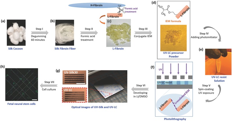
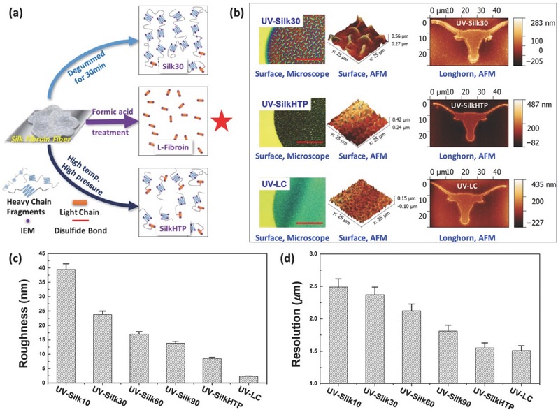
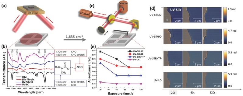
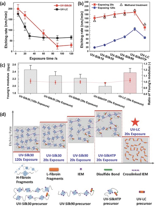
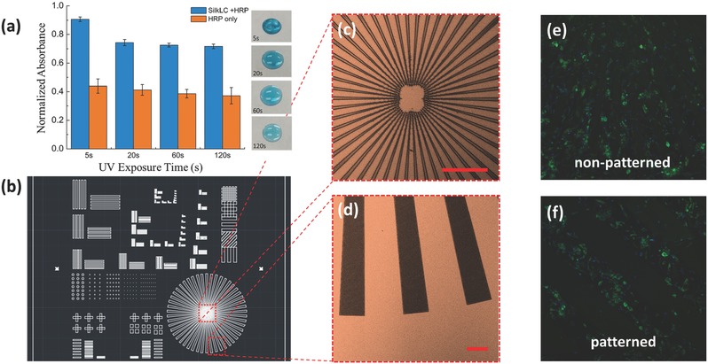
Precise patterning of biomaterials has widespread applications, including drug release, degradable implants, tissue engineering, and regenerative medicine. Patterning of protein-based microstructures using UV-photolithography has been demonstrated using protein as the resist material. The Achilles heel of existing protein-based biophotoresists is the inevitable wide molecular weight distribution during the protein extraction/regeneration process, hindering their practical uses in the semiconductor industry where reliability and repeatability are paramount. A wafer-scale high resolution patterning of bio-microstructures using well-defined silk fibroin light chain as the resist material is presented showing unprecedent performances. The lithographic and etching performance of silk fibroin light chain resists are evaluated systematically and the underlying mechanisms are thoroughly discussed. The micropatterned silk structures are tested as cellular substrates for the successful spatial guidance of fetal neural stems cells seeded on the patterned substrates. The enhanced patterning resolution, the improved etch resistance, and the inherent biocompatibility of such protein-based photoresist provide new opportunities in fabricating large scale biocompatible functional microstructures.
Keywords: biopatterning; protein photolithography; silk fibroin light chain.
Figures
Similar articles
-
Wafer-Scale Multilayer Fabrication for Silk Fibroin-Based Microelectronics.ACS Appl Mater Interfaces. 2019 Jan 9;11(1):115-124. doi: 10.1021/acsami.8b13170. Epub 2018 Dec 10. ACS Appl Mater Interfaces. 2019. PMID: 30480426
-
Precise patterning of silk microstructures using photolithography.Adv Mater. 2013 Nov 20;25(43):6207-12. doi: 10.1002/adma.201302823. Epub 2013 Aug 20. Adv Mater. 2013. PMID: 24038619
-
Using Wool Keratin as a Basic Resist Material to Fabricate Precise Protein Patterns.Adv Mater. 2019 Jul;31(28):e1900870. doi: 10.1002/adma.201900870. Epub 2019 May 12. Adv Mater. 2019. PMID: 31081271
-
Nonmulberry silk fibroin-based biomaterials: Impact on cell behavior regulation and tissue regeneration.Acta Biomater. 2022 Nov;153:68-84. doi: 10.1016/j.actbio.2022.09.021. Epub 2022 Sep 14. Acta Biomater. 2022. PMID: 36113722 Review.
-
Silk protein-based hydrogels: Promising advanced materials for biomedical applications.Acta Biomater. 2016 Feb;31:17-32. doi: 10.1016/j.actbio.2015.11.034. Epub 2015 Nov 18. Acta Biomater. 2016. PMID: 26602821 Review.
Cited by
-
Multi- and Gray-Scale Thermal Lithography of Silk Fibroin as Water-Developable Resist for Micro and Nanofabrication.Adv Sci (Weinh). 2024 Mar;11(12):e2303518. doi: 10.1002/advs.202303518. Epub 2024 Jan 17. Adv Sci (Weinh). 2024. PMID: 38234204 Free PMC article.
-
Lithographic Processes for the Scalable Fabrication of Micro- and Nanostructures for Biochips and Biosensors.ACS Sens. 2021 Jun 25;6(6):2002-2024. doi: 10.1021/acssensors.0c02704. Epub 2021 Apr 8. ACS Sens. 2021. PMID: 33829765 Free PMC article. Review.
-
Photo-Crosslinked Silk Fibroin for 3D Printing.Polymers (Basel). 2020 Dec 9;12(12):2936. doi: 10.3390/polym12122936. Polymers (Basel). 2020. PMID: 33316890 Free PMC article. Review.
-
Screening Small Metabolites from Cells as Multifunctional Coatings Simultaneously Improves Nanomaterial Biocompatibility and Functionality.Adv Sci (Weinh). 2018 May 23;5(7):1800341. doi: 10.1002/advs.201800341. eCollection 2018 Jul. Adv Sci (Weinh). 2018. PMID: 30027060 Free PMC article.
-
Methacrylated Silk Fibroin Additive Manufacturing of Shape Memory Constructs with Possible Application in Bone Regeneration.Gels. 2022 Dec 16;8(12):833. doi: 10.3390/gels8120833. Gels. 2022. PMID: 36547356 Free PMC article.
References
LinkOut - more resources
Full Text Sources
Other Literature Sources