Case Reports
. 2017 Sep 6:13:55-63.
doi: 10.1016/j.ymgmr.2017.08.004. eCollection 2017 Dec.

ALG9-CDG: New clinical case and review of the literature

Affiliations
Case Reports

ALG9-CDG: New clinical case and review of the literature

Kellie Davis et al. Mol Genet Metab Rep. .

Abstract

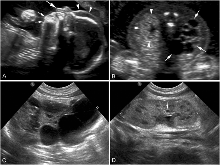
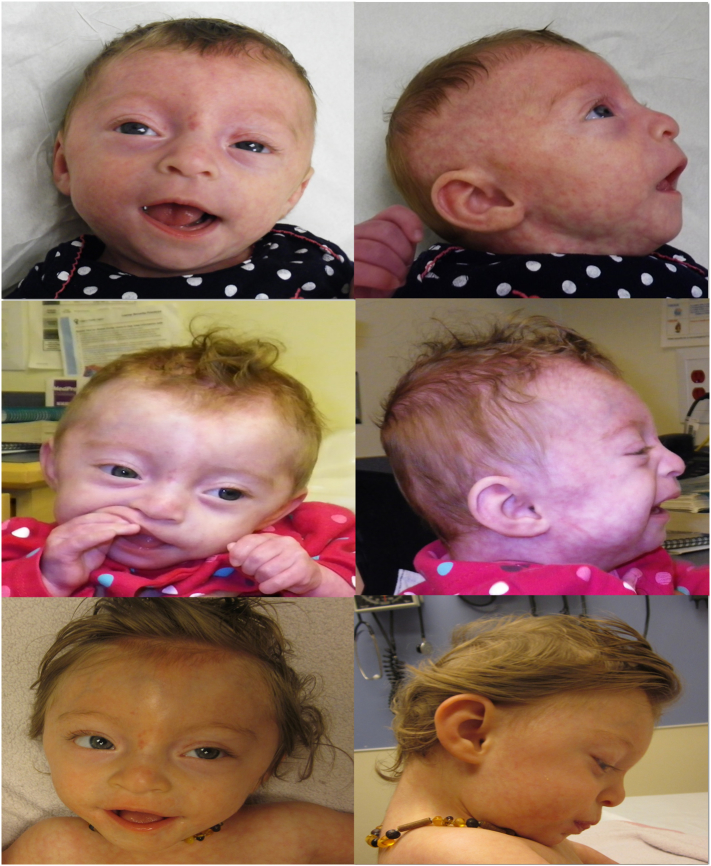
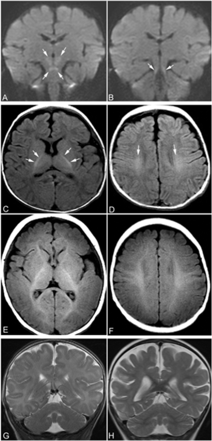
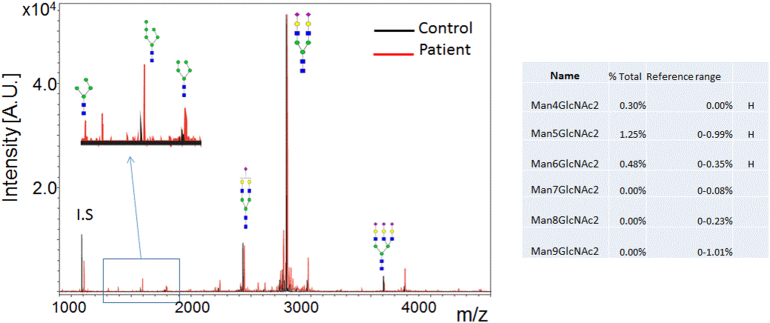
Congenital disorders of glycosylation (CDG) are a group of metabolic diseases resulting from defects in glycan synthesis or processing. The number of subgroups and their phenotypic spectrums continue to expand with most related to deficiencies of N-glycosylation. ALG9-CDG (previously CDG-IL) is the result of a mutation in ALG9. This gene encodes the enzyme alpha-1,2-mannosyltransferase. To date, a total of 10 patients from 6 different families have been reported with one of four ALG9 mutations. Seven of these patients had a similar phenotype with failure to thrive, dysmorphic features, seizures, hepatic and/or renal cysts; the other three patients died in utero from a lethal skeletal dysplasia. This report describes an additional patient with ALG9-CDG who has a milder phenotype. This patient is a term female born to Caucasian, Canadian, non-consanguineous parents of Scottish decent. Prenatally, dysmorphic features, numerous renal cysts and minor cardiac malformations were detected. Post-natally, dysmorphic features included shallow orbits, micrognathia, hypoplastic nipples, talipes equinovarus, lipodystrophy and cutis marmorata. She developed failure to thrive and seizures. The metabolic work-up included analysis of a transferrin isoelectric focusing, which showed a type 1 pattern. This was confirmed by glycan profiling, which identified ahomozygous mutation in ALG9, c.860A > G (p.Tyr287Cys) (NM_1234567890). This had been previously published as a pathogenic mutation in two Canadian patients. Our goal is to contribute to the growing body of knowledge for this disorder by describing the phenotypic spectrum and providing further insight on prognosis.

Keywords: ALG9; ALG9-CDG; CDG-IL; Congenital disorders of glycosylation; Lethal skeletal dysplasia; Transferrin isoelectrofocusing type 1 pattern.


Figures

Fig. 1
Fig. 1
Obstetrical ultrasound images from the 19 weeks' gestation anatomy scan (A, B) and post-natal renal ultrasound images obtained at day two of life (C, D). Sagittal facial profile view (A) shows prefrontal edema (arrowheads), a long philtrum (large arrow), and micrognathia (small arrows). An axial view through fetal kidneys (B) shows a markedly enlarged right kidney (arrows) in which the renal parenchyma is nearly completely replaced with simple cysts of variable sizes. The axial view also shows a morphologically normal but slightly echogenic left kidney (arrowheads). Post-natal sagittal ultrasound image of the right kidney (C) again shows a multicystic dysplastic kidney (cursors). Post-natal sagittal ultrasound image of the left kidney (D) shows morphologically normal left kidney (cursors) with a small simple cyst (arrow).
Fig. 2
Fig. 2
(left to right, top to bottom) Clinical photographs of the patient at 3, 6, and 14 months of age. Dysmorphic features shown here include hypertelorism, shallow orbits, smooth philtrum, micrognathia, fleshy ears, hypoplastic nipples and cutis marmorata. [Color figure can be viewed in the online issue, which is available at http://onlinelibrary.wiley.com/journal/10.1002/(ISSN)1552-4833.]
Fig. 3
Fig. 3
Brain MRI. At 4 months of age, an MRI of the brain shows restricted diffusion symmetrically in the superior cerebellar peduncles, midbrain, and hypothalamus (arrows in A and B). On axial T1-weighted images obtained at 4 months of age (C and D), myelination of white matter was only seen in the posterior limbs of internal capsule (arrows in C) and small areas of corona radiata (arrows in D). The myelinated white matter is hyperintense relative to the unmyelinated white matter. At 4 months of age, myelination of white matter should be seen in anterior limbs of internal capsule, optic radiations, splenium of corpus callosum, and subcortical white matter in peri-rolandic regions, as shown in the T1-weighted MR images of a normal subject obtained at the same age (E and F). The delayed myelination of the patient is easily appreciated by compared the patient's T1-weighted images to those images of the normal subject. The coronal T2-weighed images of the patient obtained at 4 months of age (G) does not show cerebral or cerebellar atrophy. However, coronal T2-weighted images at the same level obtained at 25 months of age (H) shows signs of mild to moderate global cerebral atrophy including dilatation of lateral ventricles, prominent subarachnoid spaces, and including widened cerebral sulci. No cerebellar atrophy was present on either of the MRI scans.
Fig. 4
Fig. 4
N-glycan profile in plasma for a normal control (left) and this patient (right) showing elevations in species with 4, 5, and 6 mannose units, consistent with dysfunction of alpha-1,2-mannosyltransferase. [Color figure can be viewed in the online issue, which is available at http://onlinelibrary.wiley.com/journal/10.1002/(ISSN)1552-4833.]
Fig. 5
Fig. 5
N-glycan profile in fibroblasts for a normal control (left), this patient (center) and a second patient with ALG9-CDG reported by Vleugels et al. (right) showing elevations in species with 4, 5, and 6 mannose units, consistent with dysfunction of alpha-1,2-mannosyltransferase. [Color figure can be viewed in the online issue, which is available at http://onlinelibrary.wiley.com/journal/10.1002/(ISSN)1552-4833.]
Fig. 6
Fig. 6
The top panel displays the locations of the two missense mutations within the predicted protein structure of the ALG9 protein. Both mutated residues are exposed to the ER lumen rather than residing in the cytoplasmic or predicted transmembrane domains. While the mutations are not located close together in the linear sequence of the protein, they could have close proximity in the protein tertiary structure. The bottom panel displays all three known ALG9 mutations in a linear schematic. With the limited data available thus far, there does not appear to be clustering to a particular region of the gene. The splicing mutation (c.1173 + 2T > A) causes skipping of exon 10 resulting in an out of frame transcript. [Color figure can be viewed in the online issue, which is available at http://onlinelibrary.wiley.com/journal/10.1002/(ISSN)1552-4833.]

References

    1. AlSubhi S., AlHashem A., AlAzami A., Tlili K., AlShahwan S., Lefeber D., Alkuraya F.S., Tabarki B. Further delineation of the ALG9-CDG phenotype. JIMD Rep. 2015;27:107–112. - PMC - PubMed
    1. Davids M., Kane M., He M., Wolfe L., Li X., Raihan M., Chao K., Bone W., Boerkoel C., Gahl W., Toro C. Disruption of Golgi morphology and altered protein glycosylation in PLA2G6-associated neurodegeneration. J. Med. Genet. 2016;53(3):180–189. - PMC - PubMed
    1. Eklund E.A., Freeze H.H. The congenital disorders of glycosylation: a multifaceted group of syndromes. NeuroRx. 2006 Apr;3(2):254–263. - PMC - PubMed
    1. Frank C.G., Grubenmann C.E., Eyaid W., Berger E.G., Aebi M., Hennet T. Identification and functional analysis of a defect in the human ALG9 gene: definition of congenital disorder of glycosylation type IL. Am. J. Hum. Genet. 2004;75:146–150. - PMC - PubMed
    1. Hong Z., Kajiura H., Su W., Jin H., Kimura A., Fujiyama K., Li J. Evolutionarily conserved glycan signal to degrade aberrant brassinosteroid receptors in Arabidopsis. Proc. Natl. Acad. Sci. U. S. A. 2012;109(28):11437–11442. - PMC - PubMed

Publication types

LinkOut - more resources