Review
. 2017 Aug;7(4):384-397.
doi: 10.21037/qims.2017.08.02.

Thoracic wall trauma-misdiagnosed lesions on radiographs and usefulness of ultrasound, multidetector computed tomography and magnetic resonance imaging

Affiliations
Review

Thoracic wall trauma-misdiagnosed lesions on radiographs and usefulness of ultrasound, multidetector computed tomography and magnetic resonance imaging

Xavier Tomas et al. Quant Imaging Med Surg. 2017 Aug.

Abstract

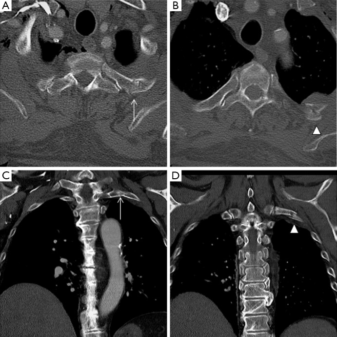
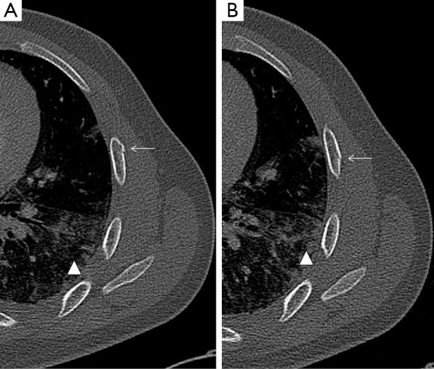
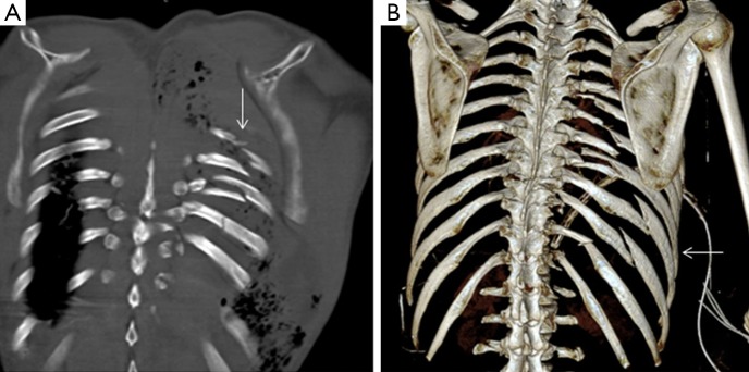
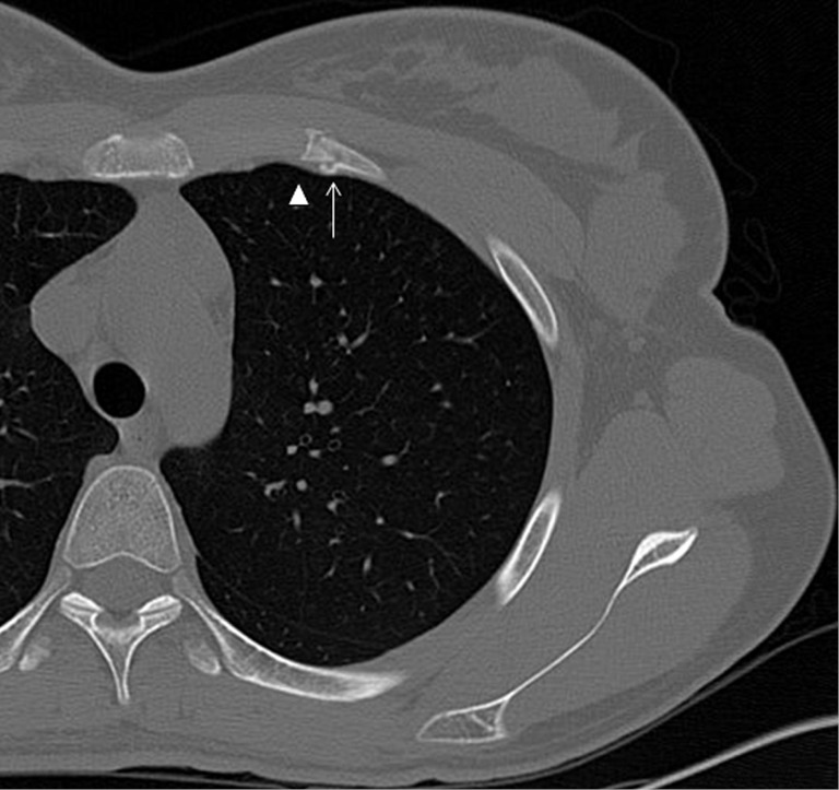
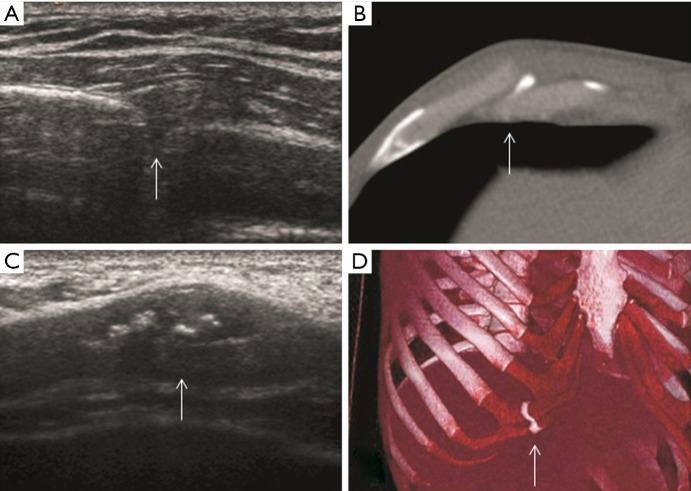
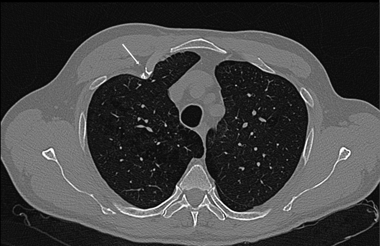
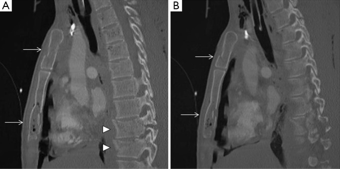
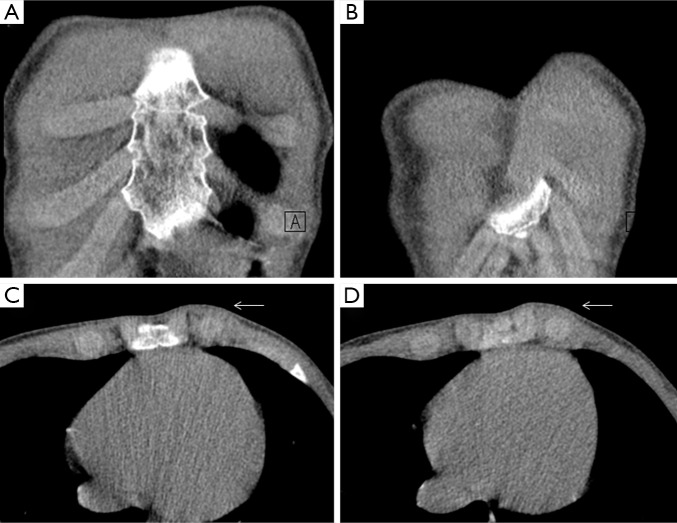
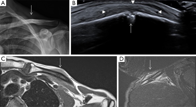
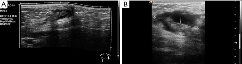
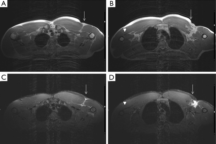
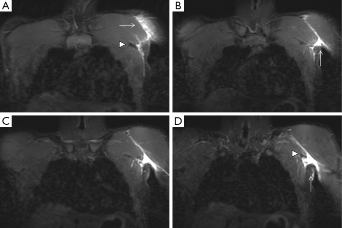
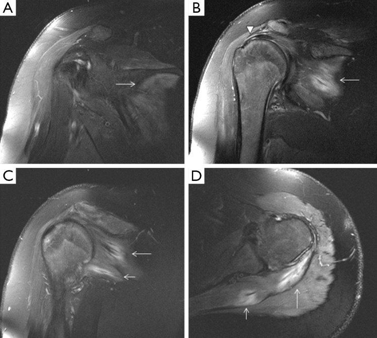
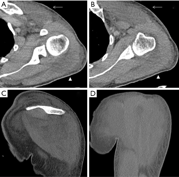
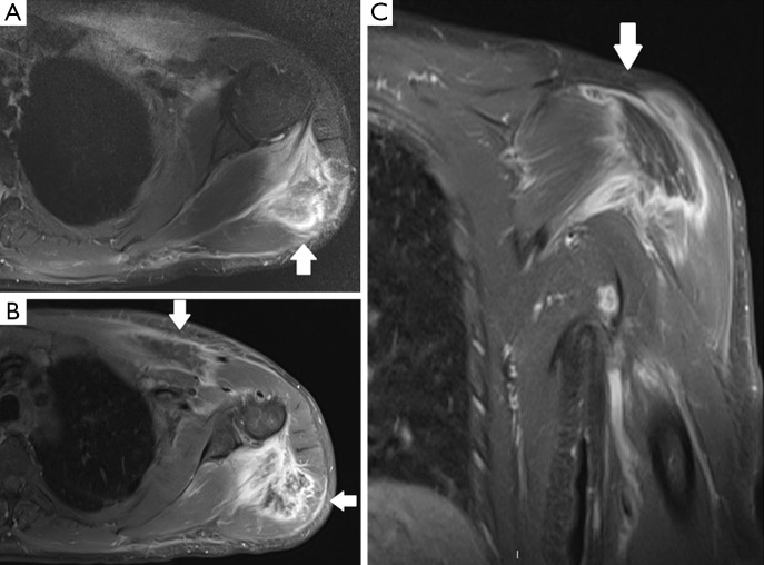
Blunt injuries to the chest wall are an important chapter on emergency room (ER) departments, being the third most common injuries in trauma patients which ominous complications could appear. This article describes different types of traumatic events affecting the chest wall, which maybe misdiagnosed with conventional X-ray. Special emphasis has been done in computed tomography (CT) and multidetector CT (MDCT) imaging. This technique is considered the "gold-standard" for those traumatic patients, due to its fast acquisition covering the whole area of interest in axial plane, reconstructing multiplanar (2D, 3D) volume-rendered images with a superb quality and angiographic CT capabilities for evaluating vascular damage. Complementary techniques such as ultrasonography (US) and magnetic resonance imaging (MRI) may improve the diagnostic accuracy due to its great capacity in visualising soft-tissue trauma (muscle-tendinous tears) and subtle fractures. All these imaging methods have an important role in quantifying the severity of chest wall trauma. The findings of this study have been exposed with cases of our archives in a didactic way.

Keywords: Magnetic resonance imaging (MRI); injuries; multidetector computed tomography (MDCT); thoracic wall; ultrasonography (US); wounds.


Conflict of interest statement

Conflicts of Interest: The authors have no conflicts of interest to declare.

Figures

Figure 1
Figure 1
Thoracic MDCT of a polytraumatic patient. (A,B) Axial and (C,D) coronal views show a non-displaced fracture of the second left rib (arrows) and a displaced fracture of the third left rib (arrowheads). MDCT, multidetector computed tomography.
Figure 2
Figure 2
Thoracic MDCT of a patient with severe cough. Axial planes highlight a buckle fracture of the rib. There is disruption of the outer cortex (arrows) with no observable fracture of the inner cortex. Patchy lung infiltrates are found (arrowheads). MDCT, multidetector computed tomography.
Figure 3
Figure 3
Thoracic MDCT of a polytraumatic patient. (A) Coronal view shows several displaced and non-displaced rib fractures, corresponding to flail chest or “volet” (arrow); (B) volume-rendered 3D image of the same patient demonstrates the presence of multiple costal fractures and flail chest (arrow). MDCT, multidetector computed tomography.
Figure 4
Figure 4
Fracture of the anterior arch of the second left rib (arrow) adjacent to the costochondral joint (arrowhead).
Figure 5
Figure 5
Costal cartilage fractures. (A) Axial ultrasound image shows a discontinuity in the echogenic line (arrow), corresponding to the anterior aspect of the hypoechogenic cartilage with a cleft, and a contiguous anterior hematoma; (B) axial MDCT image of the seventh costal arch demonstrates interruption and displacement of the cartilage (arrow); (C) axial ultrasound image in a distal rib shows swelling and a hypoechogenic area in the cartilage with focal calcifications (arrow) representing a subacute fracture; (D) volume-rendered MDCT image in a patient with a thoracic trauma three years ago shows a perpendicular ossification in the right ninth and tenth costal cartilages (arrow) related to previous fracture.
Figure 6
Figure 6
MDCT axial view manifests an asymmetry of the thoracic wall in relation to deformity in the chondrocostal joint of the second right rib (arrow). MDCT, multidetector computed tomography.
Figure 7
Figure 7
Thoracic MDCT of a polytrauma patient. (A,B) Axial and (C,D) coronal views show a subtle lucent line in the upper and left margin of the sternal manubrium (arrows), corresponding to a non-displaced fracture. Other imaging signs such a displaced rib fracture (short arrow) or soft-tissue air-bubbles can be seen (arrowheads). MDCT, multidetector computed tomography.
Figure 8
Figure 8
Thoracic MDCT of a polytrauma patient fallen from high altitude. (A,B) Sagittal view demonstrates a fracture of the manubrium and the body of sternum with several fracture lines and displacement of bone fragments (arrows). Vertebral fractures can be observed (arrowheads). MDCT, multidetector computed tomography.
Figure 9
Figure 9
MDCT. (A,B) Coronal and (C,D) axial views show an asymmetric protrusion of the left anterior chest wall, including the sternum and adjacent costal cartilages (arrows), non-related to a traumatic episode. MDCT, multidetector computed tomography.
Figure 10
Figure 10
Stress fracture of the left clavicle in a young patient after physical exertion. (A) AP radiograph of the clavicle shows irregularity and inflection in the cortical of the middle third of the clavicle (arrow); (B) oblique sonogram obtained along the clavicle shows a fracture with focal cortical disruption (arrow) surrounded by a small hematoma (arrowheads); (C) axial T1 SE-weighted MR image of the left clavicle demonstrates a low signal in the middle third with cortical disruption and periosteal reaction (arrows); (D) oblique STIR-weighted MR image shows high signal in the bone marrow and contiguous soft-tissues reflecting oedema and hematoma due to stress fracture (arrows). MR, magnetic resonance.
Figure 11
Figure 11
Patient with suspicion of rupture of the left pectoralis major muscle. (A) Sagittal ultrasound demonstrates detachment of the pectoralis major tendon from its insertion on the humerus (arrows); (B) axial ultrasound depicts a haematoma.
Figure 12
Figure 12
Axial T1-weighted (A,B) and STIR-weighted (C,D) MR images of the chest. White arrows depict soft-tissue swelling of the pectoralis major tendon, which has detached from the humerus and retracted medially. Arrowheads demonstrate the normal appearance of the right pectoralis major tendon. MR, magnetic resonance.
Figure 13
Figure 13
Coronal STIR-weighted contiguous slices (A-D) MR images of the chest. Arrows highlight the edema and hematoma present in the left pectoralis major area. Arrowheads show the detached, retracted tendon. MR, magnetic resonance.
Figure 14
Figure 14
Coronal-oblique (A-C) and axial (D) STIR-weighted MR images reflects simultaneous muscle strains of infraspinatus and teres minor muscles close to the scapula. Overall heterogeneous increase of signal intensity was depicted (arrows), revealing rupture of the muscle fibers. The supraspinatus tendon (arrowhead) remains intact. MR, magnetic resonance.
Figure 15
Figure 15
MDCT of a patient who was found unconscious, lying over his left shoulder. (A,B) Axial, and (C,D) coronal views show swelling of the left pectoral major (arrows) and deltoid muscles (arrowheads) with coarse oedema of subcutaneous tissue. MDCT, multidetector computed tomography.
Figure 16
Figure 16
Axial fat-suppressed PD-weighted (A), and fat-suppressed SET1-weighted MR images in axial (B) and coronal (C) views after intravenous administration of gadolinium of the same patient that Figure 14. Central area of pectoralis major and deltoid muscles is darker (A) and not enhancing, though the surroundings did enhance (B,C). This finding is compatible with myonecrosis due to a deep tissue injury. MR, magnetic resonance.

Similar articles

Cited by

References

    1. Kaewlai R, Avery LL, Asrani AV, Novelline RA. Multidetector CT of blunt thoracic trauma. Radiographics 2008;28:1555-70. 10.1148/rg.286085510 - DOI - PubMed
    1. The American College of Surgeons Committee on Trauma Leadership. In: Clark DE, Fantus RJ, eds. National Trauma Data Bank (NTDB) Annual Report 2007. Chicago: American College of Surgeons, 2007:1-64.
    1. Exadaktylos AK, Sclabas G, Schmid SW, Schaller B, Zimmermann H. Do we really need routine computed tomographic scanning in the primary evaluation of blunt chest trauma in patients with "normal" chest radiograph? J Trauma 2001;51:1173-6. 10.1097/00005373-200112000-00025 - DOI - PubMed
    1. Omert L, Yeaney WW, Protetch J. Efficacy of thoracic computerized tomography in blunt chest trauma. Am Surg 2001;67:660-4. - PubMed
    1. Rodriguez RM, Baumann BM, Raja AS, Langdorf MI, Anglin D, Bradley RN, Medak AJ, Mower WR, Hendey GW. Diagnostic yields, charges, and radiation dose of chest imaging in blunt trauma evaluations. Acad Emerg Med 2014;21:644-50. 10.1111/acem.12396 - DOI - PubMed

LinkOut - more resources