Review
. 2017 Aug;7(4):434-442.
doi: 10.21037/qims.2017.08.04.

Endovascular management of arterial injuries after blunt or iatrogenic renal trauma

Affiliations
Review

Endovascular management of arterial injuries after blunt or iatrogenic renal trauma

Romaric Loffroy et al. Quant Imaging Med Surg. 2017 Aug.

Abstract

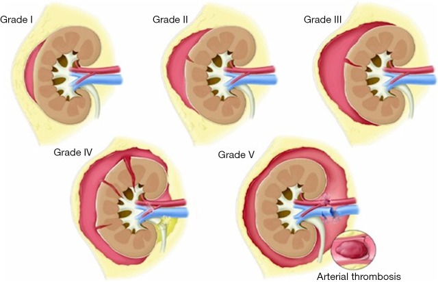
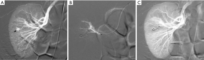
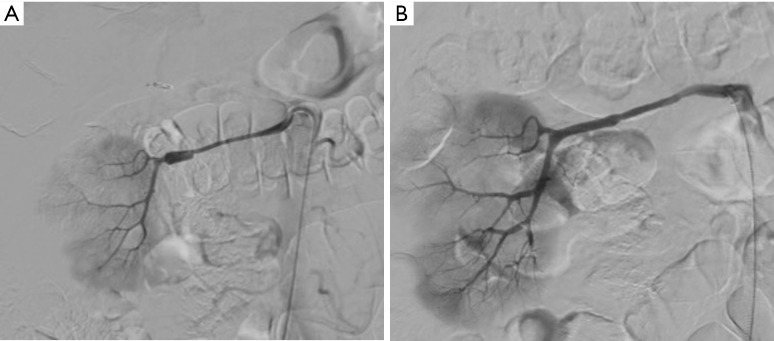
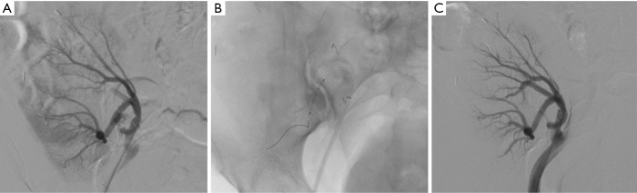
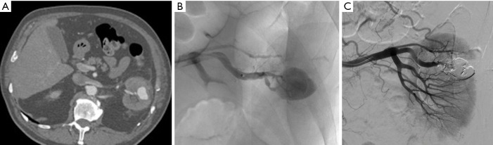
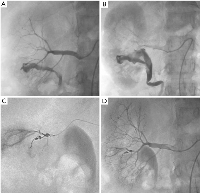
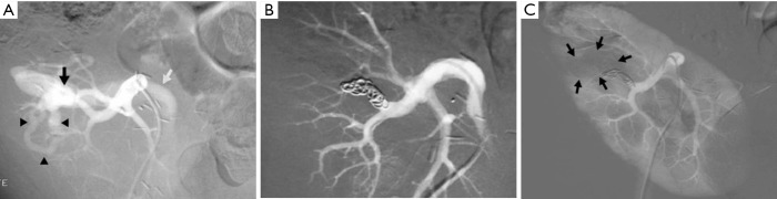
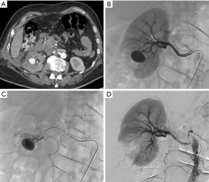
The kidney is the third most common abdominal organ to be injured in trauma, following the spleen and liver, respectively. The most commonly used classification scheme is the American Association for the Surgery of Trauma (AAST) classification of blunt renal injuries, which grades renal injury according to the size of laceration and its proximity to the renal hilum. Arteriovenous fistula and pseudoaneurysm are the most common iatrogenic biopsy-related or surgery-related vascular injuries in native kidneys. The approach to renal artery injuries has changed over time from more aggressive intervention to more conservative observational or endovascular management, including selective transcatheter arterial embolization (TAE) and the placement of stents/stent grafts. In this article, we describe the role and technical aspects of endovascular interventions in the management of arterial injuries after blunt or iatrogenic renal trauma.

Keywords: Kidney; arterial embolization; blunt trauma; iatrogenic lesion; interventional radiology.


Conflict of interest statement

Conflicts of Interest: The authors have no conflicts of interest to declare.

Figures

Figure 1
Figure 1
Kidney trauma staging.
Figure 2
Figure 2
Emergency embolization for arterial injury after blunt renal trauma in a 51-year-old woman. (A) Extravasation of contrast medium (pseudoaneurysm-like lesion) from lower distal-pole branch at selective angiography indicates continuous bleeding (arrow); (B) selective embolization of feeding artery using detachable microcoils; (C) control angiogram shows complete and selective occlusion of bleeding branch, with no active bleeding.
Figure 3
Figure 3
Late endovascular management of posttraumatic renal artery dissection in a 63-year-old man. (A) Signs of nonocclusive wall dissection of the right distal renal artery with secondary development of a false-aneurysm lesion; (B) angiogram after placement of a coronary covered stent showing normal renal artery.
Figure 4
Figure 4
Active bleeding following renal transplantation in a 46-year-old woman. (A) Selective arterial angiogram showing active extravasation from truncular part of lower-pole segmental branch of transplanted renal artery; (B) selective coronary stent-graft deployment; (C) complete cessation of active bleeding on post-procedural angiogram with conservation of the parent artery patency.
Figure 5
Figure 5
Large parenchymal false aneurysm into the left kidney after nephron-sparing surgery in a 58-year-old patient. (A) CT scan showing the false aneurysm into the left kidney; (B) superselective catheterization of the feeding artery and false aneurysm; (C) results after selective embolization of the aneurysmal sac and feeding artery with Onyx® demonstrating complete exclusion of the lesion with small cortical infarct.
Figure 6
Figure 6
Massive hematuria with hypovolemic shock 2 hours after performing percutaneous kidney biopsy in a 56-year-old woman. (A,B) Presence of high-flow arteriocaliceal fistula on emergency renal angiography with rapid opacification of urinary cavities; (C) microcoil embolization of two abnormal vessels that were responsible for hematuria; (D) complete occlusion of fistula and cessation of bleeding are seen on post-embolization angiogram.
Figure 7
Figure 7
Progressive arterial hypertension in a 43-year-old patient one year after percutaneous renal allograft biopsy. (A) Selective arterial angiogram: there is large arteriovenous fistula in upper-pole segmental branch of transplanted renal artery and pseudoaneurysm (black arrow) with marked arteriovenous shunting (arrowheads) and early venous filling (white arrow). Note absence of nephrogram; (B) control angiogram after selective embolization of afferent artery with 0.035’’ coils: complete occlusion of pedicular aneurysm and fistula, and improvement in nephrogram; (C) post-embolization angiogram (parenchymal phase): renal infarction is seen in less than 10% of renal parenchyma (arrows).
Figure 8
Figure 8
Bleeding from a pseudoaneurysm of the right kidney 3 weeks after nephron-sparing surgery. (A) CT scan showing the false aneurysm into the parenchyma; (B,C) selective and superselective angiograms of the right renal artery confirming the large vascular lesion; (D) angiography after superselective microcoil embolization of the feeding artery. Note only small parenchymal ischemia.

Similar articles

Cited by

References

    1. Titton RL, Gervais DA, Boland GW, Mueller PR. Renal trauma: radiologic evaluation and percutaneous treatment of nonvascular injuries. AJR Am J Roentgenol 2002;178:1507-11. 10.2214/ajr.178.6.1781507 - DOI - PubMed
    1. Santucci RA, McAninch JW, Safir M, Mario LA, Service S, Segal MR. Validation of the American Association for the Surgery of Trauma organ injury severity scale for the kidney. J Trauma 2001;50:195-200. 10.1097/00005373-200102000-00002 - DOI - PubMed
    1. Mani NB, Kim L. The role of interventional radiology in urologic tract trauma. Semin Intervent Radiol 2011;28:415-23. 10.1055/s-0031-1296084 - DOI - PMC - PubMed
    1. Ramaswamy RS, Darcy MD. Arterial Embolization for the Treatment of Renal Masses and Traumatic Renal Injuries. Tech Vasc Interv Radiol 2016;19:203-10. 10.1053/j.tvir.2016.06.005 - DOI - PubMed
    1. Loffroy R, Abualsaud B, Delgal A, Guiu B, Kermarrec I, Michel F, Cormier L, Mousson C, Majbri N, Rebibou JM, Cercueil JP, Krausé D. Role of percutaneous arterial embolization in renal pathology. Prog Urol 2010;20:161-71. 10.1016/j.purol.2009.07.001 - DOI - PubMed

LinkOut - more resources