Review
. 2017 Aug;7(4):443-460.
doi: 10.21037/qims.2017.07.01.

MR imaging of the traumatic triangular fibrocartilaginous complex tear

Affiliations
Review

MR imaging of the traumatic triangular fibrocartilaginous complex tear

Alex W H Ng et al. Quant Imaging Med Surg. 2017 Aug.

Abstract

Triangular fibrocartilage complex is a major stabilizer of the distal radioulnar joint (DRUJ). However, triangular fibrocartilage complex (TFCC) tear is difficult to be diagnosed on MRI for its intrinsic small and thin structure with complex anatomy. The purpose of this article is to review the anatomy of TFCC, state of art MRI imaging technique, normal appearance and features of tear on MRI according to the Palmar's classification. Atypical tear and limitations of MRI in diagnosis of TFCC tear are also discussed.

Keywords: MRI; arthrogram; tear; the triangular fibrocartilage complex (TFCC); wrist.


Conflict of interest statement

Conflicts of Interest: The authors have no conflicts of interest to declare.

Figures
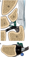
Figure 1
Figure 1
Schematic diagram shows all the TFCC components, including coronal (A) and axial (B) images. The triangular fibrocartilage or articular disc is a central component of the TFCC. On the medial side, the TFC attaches to ulnar styloid process by a distal lamina (S) and the ulnar fovea by a proximal lamina (F). On the dorsal and volar aspects, the TFC is inseparable from the dRUL and vRUL. On the radial side, the TFC attaches to the distal radial articular cartilage (R). Towards the dorsal side of the ulnar aspect, the TFC attaches to the UMH and the UCL. At around this region, the TFC also attaches to the ECU tendon sheath. On the volar aspect, the TFC attaches to the lunate (L) and triquetral (T) bones via the ulnotriquetral (UT) and ulnolunate (UL) ligaments respectively. U, distal ulna; H, hamate. TFCC, triangular fibrocartilage complex; dRUL, dorsal radioulnar ligament; vRUL, volar radioulnar ligaments; UMH, ulnomeniscal homologue; UCL, ulnar collateral ligament; ECU, extensor carpi ulnaris.
Figure 2
Figure 2
Real time situation when the patient is undergoing MRI examination of the wrist within the MRI scanner. (A) In the MRI scanner, the patient lies prone with the hand above the head in a “superman” position. Superman position allows the wrist to be scanned in the isocenter of the magnetic field, which is more homogeneous. The wrist in this position is slightly semipronated; (B) a dedicated wrist coil achieves a high resolution and signal to noise ratio. Motion artefact is the most commonly encountered problem during wrist imaging as the patient needs to maintain still for approximately 20–30 minutes in prone and superman position.
Figure 3
Figure 3
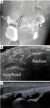
Injection of diluted gadolinium is needed to obtain a good MR arthrogram. (A) Fluoroscopic guidance is the traditional method and the needle (block arrowhead) can be punctured under real time scanning. This method is better than ultrasound guidance because flow of contrast can be seen during the injection and any communication from one compartment to another can be seen which suggests communicating tear. In this patient, contrast flow from the radiocarpal compartment to the distal radioulnar compartment through a defect in TFCC (long arrow). Contrast can also be seen flowing to the prestyloid recess (block arrow). No communication with the mid-carpal compartment; (B) ultrasound guidance of injection of diluted gadolinium into the radiocarpal joint. The needle can be identified by its echogenic linear appearance (block arrowheads). The tip can be seen contacting the distal radius; (C) after contrast injection into the both compartments, fluid distension of the radiocarpal and mid-carpal joints can be identified (asterisks). TFCC, triangular fibrocartilage complex.
Figure 4
Figure 4
The current likely hierarchy of techniques for wrist imaging with respect to depiction of tears and articular cartilage surface is suggested.
Figure 5
Figure 5
Proton density fat-suppressed MR arthrography images showing the important components of the TFCC on coronal images from volar to dorsal. (A) Ulnotriquetral (long solid arrow) and ulnolunate ligaments (short solid arrow); (B) triangular fibrocartilage articular disc (TFC) (asterisk) with radial attachment (R), foveal attachment (short block arrow), ulnar styloid process attachment (long block arrow), meniscal homologue attachment (arrowhead); (C) most dorsal aspect of TFCC with dRUL (block arrowhead) and ECU tendon sheath attachment (ECU). Small low signal foci in the ulnocarpal joint which is due to small gas locules introduced during injection of contrast. TFCC, triangular fibrocartilage complex; dRUL, dorsal radioulnar ligaments; ECU, extensor carpi ulnaris.
Figure 6
Figure 6
Normal appearance of the TFCC on sagittal T2W fat-suppressed (A) and axial proton-density fat suppressed (B) images. (A) The TFC (asterisk) is concave in appearance on a sagittal view. On the volar side, the TFC is attaching to the volar radioulnar ligament (vRUL) (long solid arrow) and more distally through the ulnotriquetral ligament (short solid arrows). On the dorsal side, the TFC is attaching to the dorsal radioulnar ligament (block block arrow). These structures are not seen as distinct structures on MRI when they are normal; (B) the TFC (asterisk) is barely seen on axial image. On the volar side, the TFC is attaching to a low signal linear structure representing the volar radioulnar ligament (vRUL). On the radial side, it attaches to the distal radial cartilaginous rim (R). On the ulnar side, the attachments are very difficult to be appreciated at this level. However, the location of the distal ulnar styloid process (short block arrow) and the ECU can be appreciated on this image. Fluid within the pre-styloid recess is seen (arrowhead). TFCC, triangular fibrocartilage complex; ECU, extensor carpi ulnaris.
Figure 7
Figure 7
Schematic diagram showing the Palmar classification of TFCC tears. (A) Coronal view shows the different types of tear correspond to the location of the tear. Type 1A is central TFC perforation. 1B, peripheral ulnar side TFCC tear (± ulna styloid fracture). 1C, distal TFCC disruption (disruption of distal UC ligaments) and 1D, radial TFCC disruption (± sigmoid notch fracture); (B) axial view shows the different tear locations. A tear on the volar and dorsal sides of the TFC involving the volar (vRUL) or dorsal radioulnar ligament (dRUL) is not included in the Palmar classification. TFCC, triangular fibrocartilage complex.
Figure 8
Figure 8
Type 1A tear. (A) Schematic drawing showing the tear (pink circle) at the central or paracentral part of the TFCC. Proton density fat suppression Coronal MRI images showing (B) full thickness tear with a small gap filled with fluid (short solid arrow). There is a small remnant of TFC at the radial attachment (long block arrow). A small subchondral cyst is at the proximal ulnar side of the lunate bone (asterisk); (C) partial thickness tear at the undersurface of the TFC (short block arrow). The distal surface of TFC is intact with the contour preserved (solid arrowhead); (D) contour irregularity (block arrowhead) of the TFC is also a sign of TFC tear as in this case which was confirmed to be a communicating full thickness tear during arthroscopy. TFCC, triangular fibrocartilage complex.
Figure 9
Figure 9
Type 1B tear. (A) Schematic drawing showing the tear (pink circles) at the proximal and distal laminae of TFCC which attach to the ulnar fovea and distal ulnar styloid process respectively. Proton-density fat-suppressed coronal MRI images showing (B) full-thickness tear as evident by complete loss of fibres of proximal (short solid arrow) which are expected to attach to the fovea and the distal ulnar styloid process; (C) another patient with a full thickness tear at the proximal (solid arrowhead) and distal laminae (block arrowhead) as evidenced by fibre discontinuity. During arthroscopy, a hook sign and trampoline effect are suggestive of unstable tear of the TFCC. Trampoline sign is positive when TFCC is soft and lax while hook test is positive when the TFCC can be pulled upward and radially towards the center during the arthroscopy using the probe. They are indicative of peripheral tear and foveal tear respectively; (D) cystic changes within the proximal and distal laminae of the TFCC attachments (long solid arrows) due to intrasubstance partial tear; (E) oedematous and thickened fibres of the proximal and distal lamina (long block arrows) due to high grade partial tear. Overall lamina continuity seems to be maintained. TFCC, triangular fibrocartilage complex.
Figure 10
Figure 10
Type 1B tear. (A) Injury one week prior. Proton-density coronal MRI image showing moderately displaced fracture base of ulnar styloid process (short solid arrow). The distal lamina is partially torn (short block arrow) with a complete tear of the proximal lamina (solid arrowhead); (B) frontal radiograph shows fracture of ulnar styloid process (block arrowhead). The TFCC cannot be evaluated radiographically; (C) proton density fat-suppressed coronal MRI image shows avulsion fracture of distal ulnar styloid process (long solid arrow). The distal laminar fibres are intact (long block arrow) though mild increased abnormal signal suggests mild sprain. A complete tear of the proximal lamina fibres (curved solid arrow) is also present. TFCC, triangular fibrocartilage complex.
Figure 11
Figure 11
Type 1C tear. (A) Schematic drawing showing tear of the ulnolunate and ulnotriquetral ligaments (pink circles); (B) proton density fat suppressed coronal MRI image shows severe oedematous change with thickening at the ulnotriquetral ligament (short solid arrow) consistent with a intrasubstance partial tear of this ligament; (C) proton-density fat suppression coronal MRI image of another patient showed severe oedematous change with thickening at the ulnolunate ligament (short block arrows) attaching to proximal lunate (L) consistent with an intrasubstance partial tear; (D) proton-density coronal MRI image of another patient shows avulsion fracture of proximal triquetrum (T) at the attachment of the ulnotriquetral ligament (solid arrowhead).
Figure 12
Figure 12
Type 1D tear. (A) Schematic drawing showing tear at the radial attachment (pink circle); (B) proton density fat-suppressed coronal MRI image shows an avulsion tear present at the radial attachment (short solid arrow). Peripheral retraction of the TFCC is noted (short block arrow). This patient had history of fracture distal radius with intraarticular extension (solid arrowhead); (C) proton density coronal MRI of another patient showed avulsion tear at the radial attachment (solid arrowhead) associated with peripheral retraction of the TFC leaving a gap filled up with fluid (block arrowhead). There is no remnant of the TFC at the radial attachment to suggest type 1A tear. TFCC, triangular fibrocartilage complex.
Figure 13
Figure 13
Dorsal sided TFCC tear. (A) Schematic drawing showing tear at the dorsal radioulnar ligament (pink circles); (B) sagittal T1W fat-suppressed MR arthrogram image shows a partial tear on the dorsal side of the TFC (solid arrow); (C) sagittal proton density fat suppressed coronal MRI image shows almost complete detachment of the TFCC at the dorsal radioulnar ligament (solid arrow); (D) proton density fat-suppressed coronal MRI image showing a severely oedematous and thickened dorsal radioulnar ligament (solid arrowheads) due to severe partial tear; (E) T2-weighted fat suppressed sagittal MRI image of another patient showing moderate thickening and oedema of the dorsal radioulnar ligament due to a severe partial tear (block arrowheads). TFCC, triangular fibrocartilage complex.
Figure 14
Figure 14
Volar sided TFCC tear. (A) Schematic drawing depicting a tear of the volar radioulnar ligament (pink circles); (B) T1-weighted fat-suppressed axial MR arthrogram shows thickened, oedematous severely torn volar radioulnar ligament (short solid arrows); (C) corresponding sagittal T1-weighted fat suppressed MR arthrogram shows this thickened and oedematous volar radioulnar ligament (long solid arrows) is still attached to the TFC which itself is also thickened and oedematous consistent with severe partial tear (block arrows). P, pisiform; T, triquetral. ECU, extensor carpi ulnaris; TFCC, triangular fibrocartilage complex.
Figure 15
Figure 15
Ulnomeniscal homologue tear. (A) Schematic drawing depicting a tear at the ulnomeniscal homologue (pink circle); (B) coronal T1-weighted fat-suppressed MR arthrogram showing partial tear at the ulnomeniscal homologue (solid arrows) extending from the distal ulnar attachment proximally to the triquetral attachment distally. The detachment of the ulnomeniscal homologue to the periphery of TFC (asterisk) can be appreciated. TFCC, triangular fibrocartilage complex.
Figure 16
Figure 16
Atypical TFCC tear. (A) Coronal proton density fat-suppressed MR image shows atypical tear of the TFC centrally located and extending from the distal surface (block arrow) to the foveal attachment (solid arrow). This is due to a combination of Palmar classification type 1A and type 1B tears; (B) coronal proton-density fat suppressed MR image shows chronic avulsion fracture at the base of distal ulnar styloid process (solid long arrow). Associated with abnormal high signal in the attachment of TFCC (solid arrowhead). Features are compatible with Type 1B tear. There is also a small linear tear at the paracentral portion of the TFC (block long arrow) compatible with type 1A tear; (C) frontal radiograph of the same patient shows the non-united fracture at the base (block arrowhead) of the ulnar styloid process (type 2). Type 2 fracture is usually associated with TFCC disruption and DRUJ instability. About 65% of patients have non-union of ulnar styloid fractures; (D) coronal proton density fat-suppressed MR image in a patient with DRUJ instability clinically showing a severe partial tear of the proximal (curved solid arrow) and distal laminae (block curved arrow) at the styloid and foveal attachments respectively; (E) in the same patient more dorsally, there is a severe sprain of the dorsal radioulnar ligament with thickening and oedema (asterisk). TFCC, triangular fibrocartilage complex; DRUJ, distal radioulnar joint.
Figure 17
Figure 17
Dorsal subluxation of the DRUJ. (A) Axial proton density fat-suppressed MR image. There is dorsal subluxation of the distal ulna at the sigmoid notch associated with moderate subchondral cystic changes (block long arrows) of the radius; (B) coronal proton density fat-suppressed MR image of another patient showed an ulnar-sided tear of the TFCC (solid long arrow). There is severe degeneration of the DRUJ with osteophytes (solid curved arrow) and joint effusion due to chronic DRUJ instability. TFCC, triangular fibrocartilage complex; DRUJ, distal radioulnar joint.
Figure 18
Figure 18
Ulnocarpal impaction. (A) Coronal proton density fat-suppressed MR image shows type 1a paracentral region tear (solid arrow) associated with subchondral cystic changes on the ulnar proximal aspect of the lunate (block arrow) consistent with ulnocarpal impaction. Mild ulnar positive variance is present; (B) coronal proton density fat-suppressed MR image of another patient also shows type 1a tear at the paracentral region of the TFCC (solid arrowhead) associated with subchondral cystic changes on the ulnar proximal aspect of the lunate (block arrowhead) with mild overlying cartilage irregularity. Features consistent with ulnocarpal impaction. TFCC, triangular fibrocartilage complex.
Figure 19
Figure 19
Comparison of TFCC visualization by non-arthrogram MRI (A) and MR arthrogram (B). Before injection of contrast, the prestyloid recess (block curved arrow) is not distended and the radiocarpal joint surface of TFCC cannot be well delineated. After contrast injection into the joint, the joints are well distended including the prestyoid recess (block curved arrow) and DRUJ (solid curved arrow). The surface of the TFCC can now be well delineated (solid arrows). TFCC, triangular fibrocartilage complex; DRUJ, distal radioulnar joint.
Figure 20
Figure 20
Comparison of TFCC visualization using non-traction MRI (A) and traction MRI (B). Before traction, there is a tear depicted at the foveal attachment (block arrow) of the TFCC but the styloid attachment appears intact (solid arrow). After traction, discontinuity at the styloid attachment can be fully appreciated (solid arrow). The radiocarpal and the mid-carpal joints are widened following traction (arrowheads). TFCC, triangular fibrocartilage complex.

Similar articles

Cited by

References

    1. Yu JS, Habib PA. Normal MR imaging anatomy of the wrist and hand. Radiol Clin North Am 2006;44:569-81. 10.1016/j.rcl.2006.04.008 - DOI - PubMed
    1. Davis KW, Blankenbaker DG. Imaging the ligaments and tendons of the wrist. Semin Roentgenol 2010;45:194-217. 10.1053/j.ro.2009.12.005 - DOI - PubMed
    1. Chhabra A, Soldatos T, Thawait GK, Del Grande F, Thakkar RS, Means KR, Jr, Carrino JA. Current perspectives on the advantages of 3-T MR imaging of the wrist. Radiographics 2012;32:879-96. 10.1148/rg.323115741 - DOI - PubMed
    1. Zlatkin MB, Rosner J. MR imaging of ligaments and triangular fibrocartilage complex of the wrist. Radiol Clin North Am 2006;44:595-623 10.1016/j.rcl.2006.04.010 - DOI - PubMed
    1. Watanabe A, Souza F, Vezeridis PS, Blazar P, Yoshioka H. Ulnar-sided wrist pain. II. Clinical imaging and treatment. Skeletal Radiol 2010;39:837-57. 10.1007/s00256-009-0842-3 - DOI - PMC - PubMed

LinkOut - more resources