Review
. 2017:2017:7238672.
doi: 10.1155/2017/7238672. Epub 2017 Aug 28.

Unilateral Aplasia versus Bilateral Aplasia of the Vertebral Artery: A Review of Associated Abnormalities

Affiliations
Review

Unilateral Aplasia versus Bilateral Aplasia of the Vertebral Artery: A Review of Associated Abnormalities

L Vasović et al. Biomed Res Int. 2017.

Erratum in

Abstract

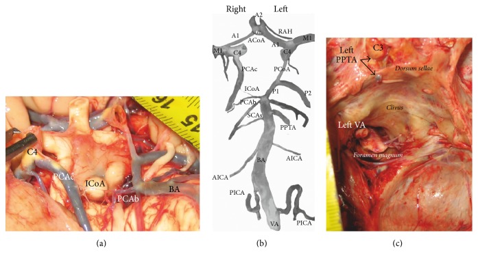
Morphological characteristics of 108 cases of uni- and bilateral aplasia of the vertebral artery (VA) in reports or images of retrospective studies, including one recent case, published between 1967 and 2016 are analyzed. Incidence, gender, persistence of carotid-vertebrobasilar anastomosis (CVBA), associated with other vascular variants, and vascular pathology in each group of uni- and bilateral VA aplasia are mutually compared. Most of the cases of VA aplasia in ages 31 to 80 were discovered in USA, Japan, and India. The bilateral VA aplasia is more common in the male gender than in the female one. The side of the VA aplasia had a significant effect on the side of CVBA persistence. Associated aplasia of other arteries was more common in cases of unilateral VA aplasia. The left VA was more commonly hypoplastic in cases of single right VA aplasia than the right VA in cases of single left VA aplasia. Aneurysms of definitive arteries were more frequent in cases of single right VA aplasia than in cases of single left VA aplasia. We claim that the aplasia of the VA probably depends on genetic factors in some races, while diseases are expressed usually in persons over 30 years of age.


Figures

Figure 1
Figure 1
Some arteries of the carotid and vertebrobasilar systems on the brain base and in the middle and posterior cranial fossae in a 77-year-old man autopsied because of myocardial infarction in the Institute of Forensic Medicine; the approval for coauthor's (MT) investigation of cadaveric cases was obtained from the Research Ethics Committee (number 01-9068-4) of our Faculty of Medicine. (a) Additional vascular component, so-called intermediate communicating artery (ICoA) between the right posterior cerebral artery (PCA) of carotid (C4) origin (PCAc) and right PCA of basilar (BA) origin (PCAb) in the cerebral arterial circle marking, is separately shown. (b) Main arteries of the carotid and vertebrobasilar systems from original picture are extracted and marked. The cerebral arterial circle has the shape of a decagon; its vascular components are as follows: subparts of the cerebral parts (C4) of paired internal carotid arteries, precommunicating part (A1) of paired anterior cerebral arteries connected by network configuration of the anterior communicating artery (ACoA), and then the left posterior communicating artery (PCoA), which connects and divides ipsilateral PCA in the precommunicating (P1) and postcommunicating (P2) parts. So-called ICoA connecting the right PCAc and PCAb presents the tenth vascular component in the cerebral arterial circle. Additional BA branch on the left side, located below the superior cerebellar artery (SCA), as a persistent primitive trigeminal artery (PPTA) is marked. Besides Heubner's artery (RAH), a large side branch of the left anterior cerebral artery and sphenoidal part (M1) of paired middle cerebral arteries are also marked. The right SCA, as partially duplicated vessel and single left SCA, as well as single anterior inferior cerebellar artery (AICA) on both sides and the right posterior inferior cerebellar artery (PICA) are side branches of the BA, while the left PICA is a branch of ipsilateral vertebral artery (VA). (c) View on a part of the middle and posterior cranial fossae of the same case. It shows the left PPTA as a branch of the cavernous part (C3) of the internal carotid artery and only the left VA in the course through the foramen magnum.

Similar articles

Cited by

References

    1. Padget D. H. Designation of the embryonic intersegmental arteries in reference to the vertebral artery and subclavian stem. The Anatomical Record. 1954;119(3):349–356. doi: 10.1002/ar.1091190306. - DOI - PubMed
    1. Effmann E. L., Whitman S. A., Smith B. R. Aortic arch development. RadioGraphics. 1986;6(6):1065–1089. doi: 10.1148/radiographics.6.6.3685519. - DOI - PubMed
    1. Vasović L., Radenković G., Trandafilović M., G. Đorđević Variable left and/or right vertebral artery in prevertebral part: a review of features in the postnatal period. Series: Medicine and Biolology. 2015;17(1):1–25.
    1. Okahara M., Kiyosue H., Mori H., Tanoue S., Sainou M., Nagatomi H. Anatomic variations of the cerebral arteries and their embryology: A pictorial review. European Radiology. 2002;12(10):2548–2561. doi: 10.1007/s00330-001-1286-x. - DOI - PubMed
    1. Vasović L., Milenković Z., Jovanović I., Čukuranović R., Jovanović P., Stefanović I. Hypoglossal artery: A review of normal and pathological features. Neurosurgical Review. 2008;31(4):385–395. doi: 10.1007/s10143-008-0145-5. - DOI - PubMed

LinkOut - more resources