. 2017 Sep 13;2(5):e00248-17.
doi: 10.1128/mSphere.00248-17. eCollection 2017 Sep-Oct.

14-3-3 Regulates Actin Filament Formation in the Deep-Branching Eukaryote Giardia lamblia

Affiliations

14-3-3 Regulates Actin Filament Formation in the Deep-Branching Eukaryote Giardia lamblia

Jana Krtková et al. mSphere. .

Abstract

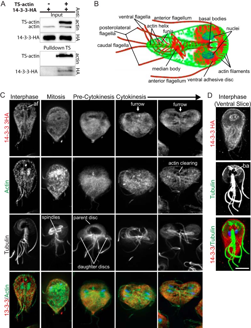
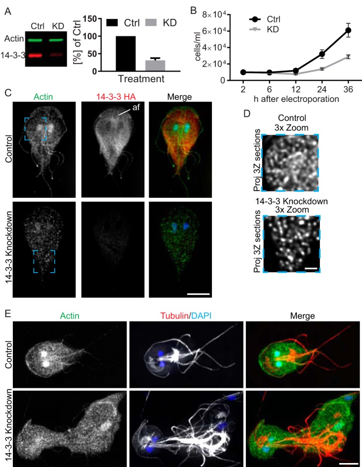
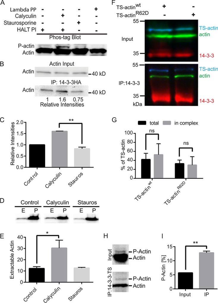
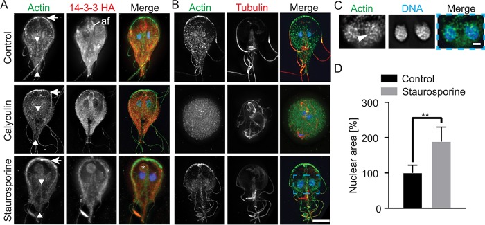
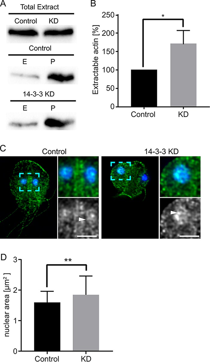
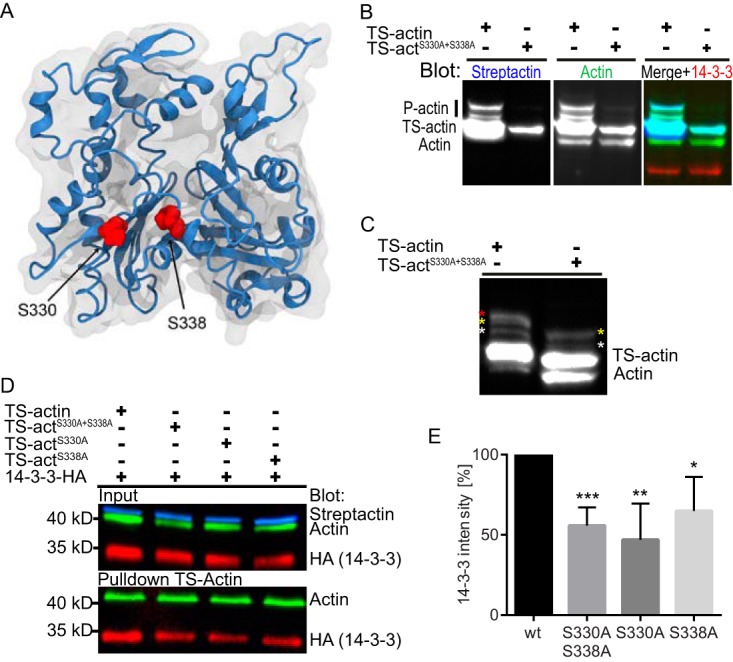
The phosphoserine/phosphothreonine-binding protein 14-3-3 is known to regulate actin; this function has been previously attributed to sequestration of phosphorylated cofilin. 14-3-3 was identified as an actin-associated protein in the deep-branching eukaryote Giardia lamblia; however, Giardia lacks cofilin and all other canonical actin-binding proteins (ABPs). Thus, the role of G. lamblia 14-3-3 (Gl-14-3-3) in actin regulation was unknown. Gl-14-3-3 depletion resulted in an overall disruption of actin organization characterized by ectopically distributed short actin filaments. Using phosphatase and kinase inhibitors, we demonstrated that actin phosphorylation correlated with destabilization of the actin network and increased complex formation with 14-3-3, while blocking actin phosphorylation stabilized actin filaments and attenuated complex formation. Giardia's sole Rho family GTPase, Gl-Rac, modulates Gl-14-3-3's association with actin, providing the first connection between Gl-Rac and the actin cytoskeleton in Giardia. Giardia actin (Gl-actin) contains two putative 14-3-3 binding motifs, one of which (S330) is conserved in mammalian actin. Mutation of these sites reduced, but did not completely disrupt, the association with 14-3-3. Native gels and overlay assays indicate that intermediate proteins are required to support complex formation between 14-3-3 and actin. Overall, our results support a role for 14-3-3 as a regulator of actin; however, the presence of multiple 14-3-3-actin complexes suggests a more complex regulatory relationship than might be expected for a minimalistic parasite. IMPORTANCEGiardia lacks canonical actin-binding proteins. Gl-14-3-3 was identified as an actin interactor, but the significance of this interaction was unknown. Loss of Gl-14-3-3 results in ectopic short actin filaments, indicating that Gl-14-3-3 is an important regulator of the actin cytoskeleton in Giardia. Drug studies indicate that Gl-14-3-3 complex formation is in part phospho-regulated. We demonstrate that complex formation is downstream of Giardia's sole Rho family GTPase, Gl-Rac. This result provides the first mechanistic connection between Gl-Rac and Gl-actin in Giardia. Native gels and overlay assays indicate intermediate proteins are required to support the interaction between Gl-14-3-3 and Gl-actin, suggesting that Gl-14-3-3 is regulating multiple Gl-actin complexes.

Keywords: 14-3-3; Rho GTPase; actin; evolutionary cell biology.


Figures

FIG 1
FIG 1
14-3-3 is associated with monomeric actin. (A) Pulldown of TS-actin demonstrating that 14-3-3 interacts with monomeric actin. (B) Diagram of actin (green) and tubulin (red) cytoskeletal structures found in interphase Giardia trophozoites. (C) Gl-14-3-3–HA (red), Gl-actin (green), tubulin (grayscale), and DNA (blue) localized in interphase, mitosis, and cytokinesis. Gl-14-3-3–HA was enriched along the intracytoplasmic portion of the anterior flagella (af). (D) Gl-14-3-3–HA (red), tubulin (green), and DNA (blue) projection spanning the ventral region only. Note Gl-14-3-3–HA in the microtubule bare area (ba) of the ventral disc (conduit for membrane trafficking). Scale bar, 5µm.
FIG 2
FIG 2
14-3-3 is required for Giardia actin cytoskeletal organization and growth. (A) Multiplexed immunoblot showing typical Gl-14-3-3–HA reduction 24 h after morpholino treatment and quantification of three independent experiments. (B) Growth curves of control (Ctrl) and Gl-14-3-3-depleted cell cultures indicate that Gl-14-3-3 is critical for Giardia culture growth (Error bars represent standard deviation [SD].) (C) Immunofluorescence staining of control and Gl-14-3-3-depleted cells scaled equally. Note enrichment of Gl-14-3-3–HA along the intracytoplasmic axonemes of the anterior flagella (af) and that depletion of Gl-14-3-3 altered actin organization. Scale bar, 5 µm. (D) Magnified view of the blue box in panel C optimally scaled to show actin filaments in the control and Gl-14-3-3-depleted cells. The puncta in Gl-14-3-3-depleted cells are short filaments; see Movie S1 for an entire image stack. Scale bar, 1 µm. (E) Actin (green) and tubulin (red) staining shows 14-3-3-depleted cells lose cell polarity and have cytokinesis defects. See Fig. S3 for further examples of knockdown phenotypes. Scale bar, 5 µm.
FIG 3
FIG 3
14-3-3–actin complex formation is phospho-dependent. (A) Immunoblot after Phos-tag phosphate-affinity electrophoresis. Cells were pretreated with DMSO or inhibitors and then HALT phosphatase inhibitor (HALT PI) was added at lysis to preserve the phosphorylation state. Calyculin A treatment increased phosphorylated-actin (P-actin) levels, and the kinase inhibitor staurosporine reduced P-actin. Phospho-isoforms were removed after lambda protein phosphatase (PP) treatment. (B) Immunoprecipitation of Gl-14-3-3–HA after calyculin A treatment led to increased actin interaction, while staurosporine treatment reduced the association of actin with Gl-14-3-3–HA. (C) Mean values of three independent experiments. Error bars represent SD. **, P < 0.01. (D) Detergent-extractable actin is increased by calyculin A treatment. E, extracted, predominantly G-actin; P, cell pellet/nonextracted, predominantly F-actin. (E) Plots are mean percentages of extractable actin from three independent experiments. Error bars represent SD. *, P < 0.05. (F) Pulldown of 14-3-3 in cells expressing wild-type TS-actin or the polymerization-defective R62D mutant. (G) Graph showing binding of wild-type TS-actin compared to the R62D polymerization-defective mutant in three independent experiments. ns, not statistically significant. (H) Compared with input, eluted protein from 14-3-3–TS pulldown shows enrichment of P-actin. (I) Quantification of three independent experiments. **, P < 0.01.
FIG 4
FIG 4
Filamentous actin structures are depleted by calyculin A treatment and enhanced by staurosporine treatment. (A) Projected images of actin (green), Gl-14-3-3–HA (red), and DNA (blue) in the presence of calyculin A and staurosporine. Arrows mark the anterior of the cell where actin intensity is reduced by calyculin A treatment (increased phosphorylation) but enhanced by staurosporine treatment (reduced phosphorylation). Arrowheads mark the intracytoplasmic caudal flagella axonemes, which are typically associated with actin. Note that calyculin A treatment resulted in loss of actin association with this structure, while staurosporine treatment increased actin association. Calyculin A treatment enriched Gl-14-3-3–HA along the intracytoplasmic axoneme of the anterior flagella (af). An asterisk marks the aberrant structure found in 30% of staurosporine-treated cells. Note this structure is associated with the bare region of the disc, a conduit of cellular trafficking. (B) Projected images of actin (green), tubulin (red), and DNA (blue) in the presence of calyculin A and staurosporine. Note that 27% of calyculin A-treated cells lost cytoskeletal organization and adopted a rounded cell shape. Scale bar, 5 µm. Nuclear area increased after staurosporine treatment. (C) A single optical section enlarged from the blue box in panel B showing actin filaments associated with the nuclei. An arrowhead marks a prominent filament. (D) Nuclear area quantified after treatment with staurosporine (control [DMSO], n = 22; staurosporine, n = 64). Values are means ± SD. **, P < 0.01. Scale bar, 1 µm.
FIG 5
FIG 5
14-3-3 knockdown alters actin extractability and increases nuclear size. (A) Detergent-extractable actin is increased by 14-3-3 knockdown (KD). E, extracted; P, cell pellet/nonextracted. (B) The plot is normalized to extracted actin from the matching control; results are from three independent experiments. Error bars represent SD. *, P < 0.05. (C) Actin (green) and DAPI (blue) staining from control and 14-3-3 KD. Nuclear size is variable in 14-3-3 KD cells. A magnified view (dashed cyan box) shows that although filaments are smaller in the cytoplasm, the large nuclei have robust filaments that span the width of the nuclei. (The white arrowhead points to the nuclear actin filament, which is also apparent in Fig. 2E.) Scale bar, 2 µm for magnified views. (D) Quantification of nuclear area indicates a 16.4% average increase in 14-3-3 KD cells compared to controls (control, n = 60; KD, n = 96). **, P < 0.01.
FIG 6
FIG 6
Rac signaling modulates 14-3-3–actin complex formation. (A) Actin filaments (green) are more prominent in tet-induced HA-RacCA-expressing cells. Note that the tet promoter is leaky and some expression is detected in uninduced control cells (images scaled equally). (B) Immunoprecipitation of actin with 14-3-3–VSVG from uninduced (−) and induced (+) HA-RacCA cell lines and quantification of actin binding from three independent experiments. ***, P < 0.001. Scale bar, 5 µm.
FIG 7
FIG 7
S330 and S338 of Gl-actin contribute to 14-3-3 complex formation. (A) Model of Gl-actin showing the positions of S330 and S338 in an actin monomer. (B) Multiplexed immunoblot of total Giardia extracts after Phos-tag phosphate-affinity electrophoresis comparing phosphorylation of TS-actin and TS-actinS330A S338A; anti-Gl-actin (green), StrepTactin-HRP (blue), and anti-HA (red). Note equal loading as indicated by 14-3-3 levels. (C) Samples from panel B were overloaded and probed with anti-Gl-actin antibody. Colored asterisks mark specific P-actin bands: note that only two bands are visible in the TS-actinS330A S338A double mutant. (D) Affinity pulldown of TS-actin variants blotted for Gl-actin and 14-3-3–HA. (E) Quantification of three independent affinity pulldown experiments shows S330 and S338 contribute to 14-3-3 association. *, P < 0.05; **, P < 0.01; ***, P < 0.001.
FIG 8
FIG 8
14-3-3–actin complex formation requires intermediate proteins. An immunoblot of affinity-purified 6×His-tagged Gl-actin (His-actin) or mock purification from wild-type trophozoites was assessed by overlay with recombinant GST–Gl-14-3-3 or the 14-3-3 binding-defective mutant GST-K53E. Interaction of GST–Gl-14-3-3 with actin and copurified proteins was revealed by incubation with anti-GST–HRP. The same membrane was stripped and probed with mouse anti-Gl-actin and again with mouse MAb anti-pSer. Silver-stained protein purifications are shown in the last inset. Molecular mass markers (kilodaltons) are on the left. The position of His-actin is indicated on the right. (B) Affinity-purified TS-actin was run on SDS-PAGE and native PAGE. The native gel analysis includes DSP-cross-linked samples to preserve native complexes. Note that DSP treatment reduces the amount of ~100-kDa dimeric 14-3-3 (red arrowhead) and increases the high-molecular-mass smear. The position of putative monomeric TS-actin (45.2-kDa expected size) is marked with a green arrowhead.

Similar articles

Cited by

References

    1. Gardino AK, Yaffe MB. 2011. 14-3-3 proteins as signaling integration points for cell cycle control and apoptosis. Semin Cell Dev Biol 22:688–695. doi:10.1016/j.semcdb.2011.09.008. - DOI - PMC - PubMed
    1. Sluchanko NN, Gusev NB. 2010. 14-3-3 proteins and regulation of cytoskeleton. Biochemistry (Mosc) 75:1528–1546. doi:10.1134/S0006297910130031. - DOI - PubMed
    1. Tzivion G, Avruch J. 2002. 14-3-3 proteins: active cofactors in cellular regulation by serine/threonine phosphorylation. J Biol Chem 277:3061–3064. doi:10.1074/jbc.R100059200. - DOI - PubMed
    1. Gohla A, Bokoch GM. 2002. 14-3-3 regulates actin dynamics by stabilizing phosphorylated cofilin. Curr Biol 12:1704–1710. doi:10.1016/S0960-9822(02)01184-3. - DOI - PubMed
    1. Kakinuma N, Roy BC, Zhu Y, Wang Y, Kiyama R. 2008. Kank regulates Rhoa-dependent formation of actin stress fibers and cell migration via 14-3-3 in Pi3k-Akt signaling. J Cell Biol 181:537–549. doi:10.1083/jcb.200707022. - DOI - PMC - PubMed

LinkOut - more resources