

Integration of digital dental casts in cone beam computed tomography scans-a clinical validation study
- PMID: 28932947
- PMCID: PMC5866842
- DOI: 10.1007/s00784-017-2203-2
Integration of digital dental casts in cone beam computed tomography scans-a clinical validation study
Abstract
Objectives: Images derived from cone beam computed tomography (CBCT) scans lack detailed information on the dentition and interocclusal relationships needed for proper surgical planning and production of surgical splints. To get a proper representation of the dentition, integration of a digital dental model into the CBCT scan is necessary. The aim of this study was to validate a simplified protocol to integrate digital dental models into CBCT scans using only one scan.
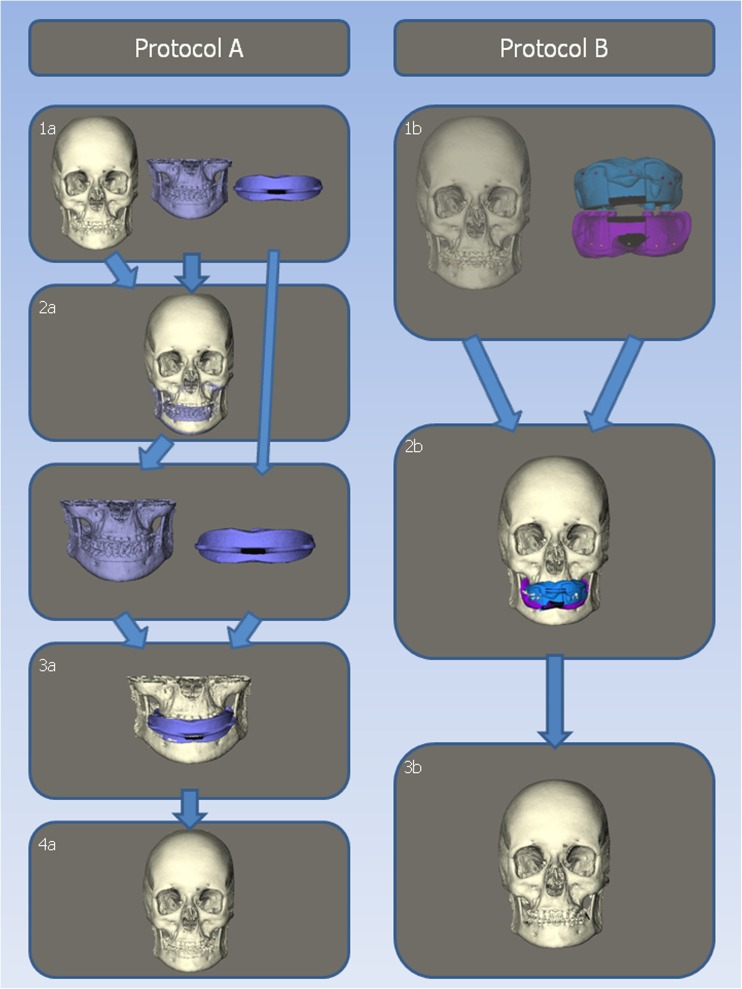
Materials and methods: Conventional protocol A used one combined upper and lower impression and two CBCT scans. The new protocol B included placement of ten markers on the gingiva, one CBCT scan, and two separate impressions of the upper and lower dentition. Twenty consecutive patients, scheduled for mandibular advancement surgery, were included. To validate protocol B, 3-dimensional reconstructions were made, which were compared by calculating the mean intersurface distances obtained with both protocols.
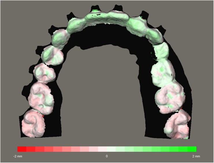
Results: The mean distance for all patients for the upper jaw is 0.39 mm and for the lower jaw is 0.30 mm. For ten out of 20 patients, all distances were less than 1 mm. For the other ten patients, all distances were less than 2 mm.
Conclusions: Mean distances of 0.39 and 0.30 mm are clinically acceptable and comparable to other studies; therefore, this new protocol is clinically accurate.
Clinical relevance: This new protocol seems to be clinically accurate. It is less time consuming, gives less radiation exposure for the patient, and has a lower risk for positional errors of the impressions compared to other integration protocols.
Keywords: Computer-assisted; Digital dental casts; Imaging; Oral and maxillofacial surgery; Orthodontics; Three-dimensional; Three-dimensional imaging.
Conflict of interest statement
Conflict of interest
The authors declare that they have no conflict of interest.
Ethical approval
All procedures performed in studies involving human participants were in accordance with the ethical standards of the institutional and/or national research committee and with the 1964 Helsinki declaration and its later amendments or comparable ethical standards.
Informed consent
Informed consent was obtained from all individual participants included in the study.
Figures


References
-
- Swennen GR, Mollemans W, De Clercq C, Abeloos J, Lamoral P, Lippens F, Neyt N, Casselman J, Schutyser F. A cone-beam computed tomography triple scan procedure to obtain a three-dimensional augmented virtual skull model appropriate for orthognathic surgery planning. J Craniofac Surg. 2009;20(2):297–307. doi: 10.1097/SCS.0b013e3181996803. - DOI - PubMed
Publication types
MeSH terms
Grants and funding
LinkOut - more resources
Full Text Sources
Other Literature Sources
