

Extracellular vesicles: how they interact with endothelium, potentially contributing to metastatic cancer cell implants
- PMID: 28933058
- PMCID: PMC5607152
- DOI: 10.1186/s40169-017-0165-2
Extracellular vesicles: how they interact with endothelium, potentially contributing to metastatic cancer cell implants
Abstract
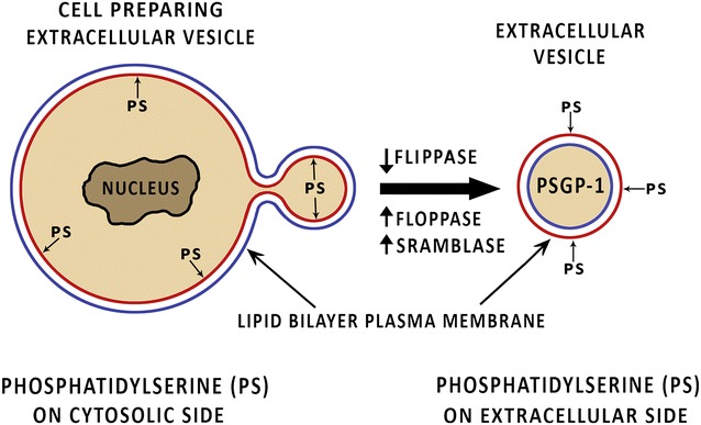
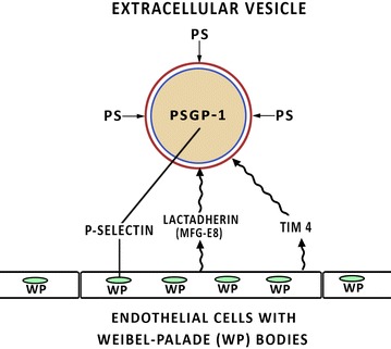
Extracellular vesicles (EV) are blebs of cellular membranes, which entrap small portions of subjacent cytosol. They are released from a variety of cells, circulate in the blood for an unknown length of time and come to rest on endothelial surfaces. They contribute to an array of physiologic pathways, the complexity of which is still being investigated. They contribute to metastatic malignant cell implants and tumor-related angiogenesis, possibly abetted by the tissue factor that they carry. It is thought that the adherence of the EV to endothelium is dependent upon a combination of their P-selectin glycoprotein ligand-1 and exposed phosphatidylserine, the latter of which is normally hidden on the inner bilayer of the intact cellular membrane. This manuscript reviews what is known about EV origins, their clearance from the circulation and how they contribute to malignant cell implants upon endothelium surfaces and subsequent tumor growth.
Keywords: Endothelium; Exosomes; Extracellular vesicles; Hypercoagulation; Metastatic carcinoma; Microparticles; P-selectin; P-selectin glycoprotein ligand 1; Tissue factor.
Figures
Similar articles
-
Extracellular Vesicles: Recent Developments in Technology and Perspectives for Cancer Liquid Biopsy.Recent Results Cancer Res. 2020;215:319-344. doi: 10.1007/978-3-030-26439-0_17. Recent Results Cancer Res. 2020. PMID: 31605237 Review.
-
Procoagulant and immunogenic properties of melanoma exosomes, microvesicles and apoptotic vesicles.Oncotarget. 2016 Aug 30;7(35):56279-56294. doi: 10.18632/oncotarget.10783. Oncotarget. 2016. PMID: 27462921 Free PMC article.
-
Extracellular vesicles and microvascular pathology: Decoding the active dialogue.Microcirculation. 2019 Feb;26(2):e12485. doi: 10.1111/micc.12485. Epub 2018 Jul 23. Microcirculation. 2019. PMID: 29923276 Review.
-
Extracellular vesicles as biomarkers in malignant pleural mesothelioma: A review.Crit Rev Oncol Hematol. 2020 Jun;150:102949. doi: 10.1016/j.critrevonc.2020.102949. Epub 2020 Apr 9. Crit Rev Oncol Hematol. 2020. PMID: 32330840 Review.
-
Extracellular Vesicles Activate a CD36-Dependent Signaling Pathway to Inhibit Microvascular Endothelial Cell Migration and Tube Formation.Arterioscler Thromb Vasc Biol. 2016 Mar;36(3):534-44. doi: 10.1161/ATVBAHA.115.307085. Epub 2016 Jan 28. Arterioscler Thromb Vasc Biol. 2016. PMID: 26821945 Free PMC article.
Cited by
-
The Potential Role of Extracellular Vesicles in COVID-19 Associated Endothelial injury and Pro-inflammation.medRxiv [Preprint]. 2020 Sep 1:2020.08.27.20182808. doi: 10.1101/2020.08.27.20182808. medRxiv. 2020. Update in: J Extracell Vesicles. 2021 Jul;10(9):e12117. doi: 10.1002/jev2.12117. PMID: 32909001 Free PMC article. Updated. Preprint.
-
Turnip Mosaic Virus Components Are Released into the Extracellular Space by Vesicles in Infected Leaves.Plant Physiol. 2019 Jul;180(3):1375-1388. doi: 10.1104/pp.19.00381. Epub 2019 Apr 24. Plant Physiol. 2019. PMID: 31019004 Free PMC article.
-
Alk5/Runx1 signaling mediated by extracellular vesicles promotes vascular repair in acute respiratory distress syndrome.Clin Transl Med. 2018 Jun 22;7(1):19. doi: 10.1186/s40169-018-0197-2. Clin Transl Med. 2018. PMID: 29931538 Free PMC article.
-
Exploring extracellular vesicles as mediators of clinical disease and vehicles for viral therapeutics: Insights from the COVID-19 pandemic.Extracell Vesicles Circ Nucl Acids. 2022;3(3):172-188. doi: 10.20517/evcna.2022.19. Epub 2022 Jul 19. Extracell Vesicles Circ Nucl Acids. 2022. PMID: 35929616 Free PMC article.
-
Detection and Investigation of Extracellular Vesicles in Serum and Urine Supernatant of Prostate Cancer Patients.Diagnostics (Basel). 2021 Mar 8;11(3):466. doi: 10.3390/diagnostics11030466. Diagnostics (Basel). 2021. PMID: 33800141 Free PMC article.
References
-
- Lötvall J, Hill AF, Hochberg F, Buzás EI, Di Vizio D, Gardiner C, et al. Minimal experimental requirements for definition of extracellular vesicles and their functions: a position statement from the International Society for Extracellular Vesicles. J Extracell Vesicles. 2014;3:26913. doi: 10.3402/jev.v3.26913. - DOI - PMC - PubMed
Publication types
LinkOut - more resources
Full Text Sources
Other Literature Sources