. 2017 Oct 2;13(4):141-155.
doi: 10.1080/15476278.2017.1358337. Epub 2017 Sep 21.

Regulation of proliferation in developing human tooth germs by MSX homeodomain proteins and cyclin-dependent kinase inhibitor p19INK4d

Affiliations

Regulation of proliferation in developing human tooth germs by MSX homeodomain proteins and cyclin-dependent kinase inhibitor p19INK4d

Darko Kero et al. Organogenesis. .

Abstract

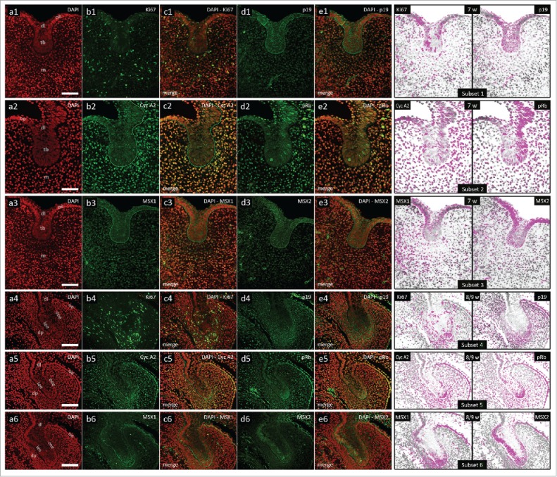
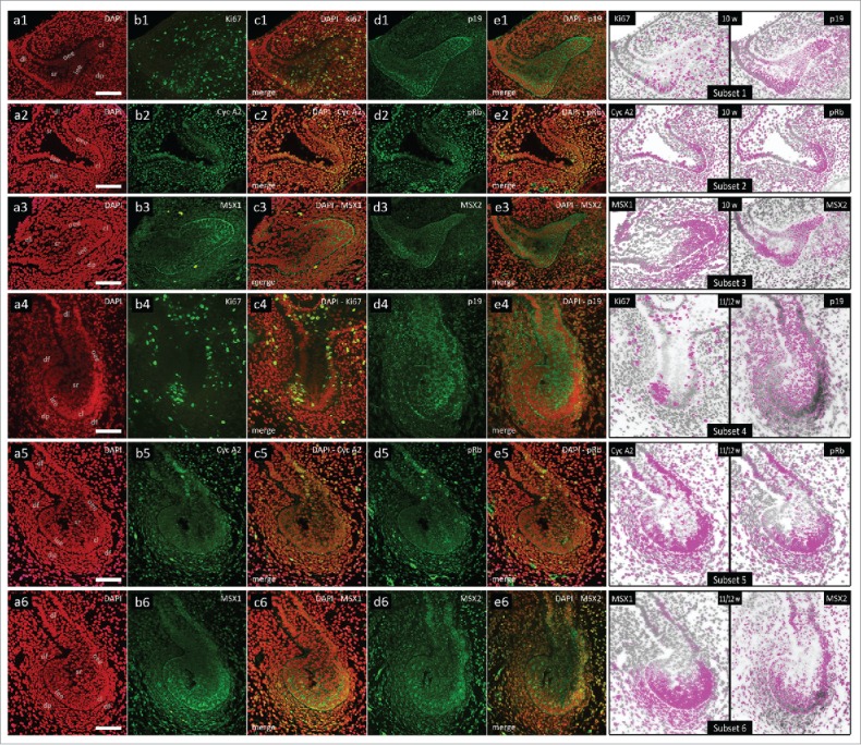
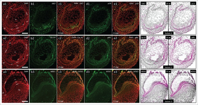
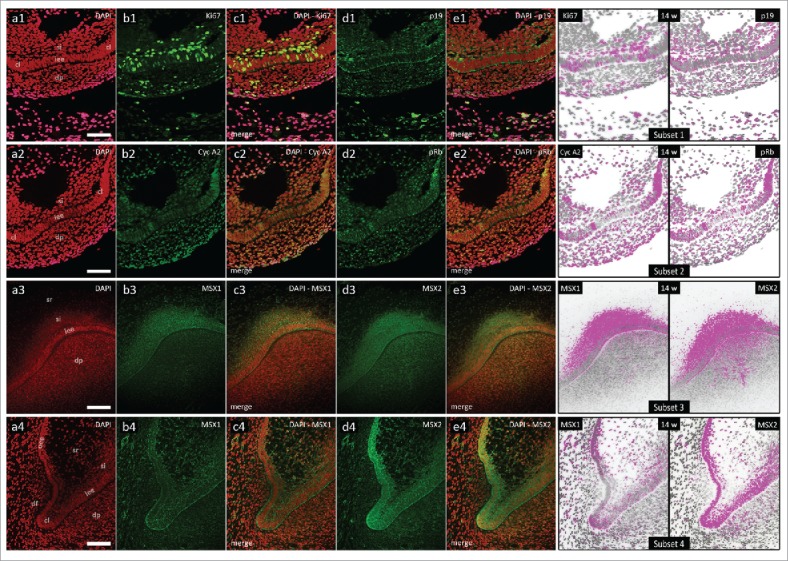
Before the secretion of hard dental tissues, tooth germs undergo several distinctive stages of development (dental lamina, bud, cap and bell). Every stage is characterized by specific proliferation patterns, which is regulated by various morphogens, growth factors and homeodomain proteins. The role of MSX homeodomain proteins in odontogenesis is rather complex. Expression domains of genes encoding for murine Msx1/2 during development are observed in tissues containing highly proliferative progenitor cells. Arrest of tooth development in Msx knockout mice can be attributed to impaired proliferation of progenitor cells. In Msx1 knockout mice, these progenitor cells start to differentiate prematurely as they strongly express cyclin-dependent kinase inhibitor p19INK4d. p19INK4d induces terminal differentiation of cells by blocking the cell cycle in mitogen-responsive G1 phase. Direct suppression of p19INK4d by Msx1 protein is, therefore, important for maintaining proliferation of progenitor cells at levels required for the normal progression of tooth development. In this study, we examined the expression patterns of MSX1, MSX2 and p19INK4d in human incisor tooth germs during the bud, cap and early bell stages of development. The distribution of expression domains of p19INK4d throughout the investigated period indicates that p19INK4d plays active role during human tooth development. Furthermore, comparison of expression domains of p19INK4d with those of MSX1, MSX2 and proliferation markers Ki67, Cyclin A2 and pRb, indicates that MSX-mediated regulation of proliferation in human tooth germs might not be executed by the mechanism similar to one described in developing tooth germs of wild-type mouse.

Keywords: MSX1; MSX2; cell cycle; development; human tooth germ; p19INK4d; proliferation.


Figures

Figure 1.
Figure 1.
Expression patterns of Ki67, p19INK4d, Cyclin A2, phosphorylated Rb, MSX1 and MSX2 in human incisor tooth germs during the bud stage and bud-to-cap stage transition. Expression patterns of p19INK4d, MSX1, MSX2 and proliferation markers Ki67, Cyclin A2 and pRb in human incisor tooth germ in the bud stage and during the bud-to-cap transition (Magnification: × 40, scale bar: 25 µm); (a1-6) DAPI staining of nuclei (inverted – red color); (b1-6, d1-6) expression patterns of investigated factors in epithelial and mesenchymal compartments of human incisor tooth germ; (c1-6, e1-6) merged image doublets of investigated factors' expression patterns with DAPI; approximation of expression domains for Ki67 and p19INK4d (Subsets 1, 4), Cyclin A2 and pRb (Subsets 2, 5), MSX1 and MSX2 (Subsets 3, 6), expression domains are displayed in magenta color (expression intensity range covered – mild to strong). Designations: oral epithelium (oe), dental lamina (dl), tooth bud (tb), jaw mesenchyme (m), outer enamel epithelium (oee), inner enamel epithelium (iee), stellate reticulum (sr), stratum intermedium (si), cervical loop (cl), dental papilla (dp).
Figure 2.
Figure 2.
Expression patterns of Ki67, p19INK4d, Cyclin A2, phosphorylated Rb, MSX1 and MSX2 in human incisor tooth germs during the cap stage. Expression patterns of p19INK4d, MSX1, MSX2 and proliferation markers Ki67, Cyclin A2 and pRb in human incisor tooth germ during the cap stage (Magnification: × 20; scale bar: 40 µm); (a1-6) DAPI staining of nuclei (inverted – red color); (b1-6, d1-6) expression patterns of investigated factors in epithelial and mesenchymal compartments of human incisor tooth germ; (c1-6, e1-6) merged image doublets of investigated factors' expression patterns with DAPI; approximation of expression domains for Ki67 and p19INK4d (Subsets 1, 4), Cyclin A2 and pRb (Subsets 2, 5), MSX1 and MSX2 (Subsets 3, 6), expression domains are dispalyed in magenta color (expression intensity range covered – mild to strong). Designations: oral epithelium (oe), dental lamina (dl), tooth bud (tb), jaw mesenchyme (m), outer enamel epithelium (oee), inner enamel epithelium (iee), stellate reticulum (sr), stratum intermedium (si), cervical loop (cl), dental papilla (dp).
Figure 3.
Figure 3.
Expression patterns of Ki67, p19INK4d, Cyclin A2, phosphorylated Rb, MSX1 and MSX2 in human incisor tooth germs during the early bell stage. Expression patterns of p19INK4d, MSX1, MSX2 and proliferation markers Ki67, Cyclin A2 and pRb in human incisor tooth germ in the early bell stage (Magnification: × 10; scale bar: 100 µm); (a1-3) DAPI staining of nuclei (inverted – red color); (b1-3, d1-3) expression patterns of investigated factors in epithelial and mesenchymal compartments of human incisor tooth germ; (c1-3, e1-3) merged image doublets of investigated factors' expression patterns with DAPI; approximation of expression domains for Ki67 and p19INK4d (Subset 1), Cyclin A2 and pRb (Subset 2), MSX1 and MSX2 (Subset 3), expression domains are displayed in magenta color (expression intensity range covered – moderate to strong). Designations: oral epithelium (oe), dental lamina (dl), tooth bud (tb), jaw mesenchyme (m), outer enamel epithelium (oee), inner enamel epithelium (iee), stellate reticulum (sr), stratum intermedium (si), cervical loop (cl), dental papilla (dp).
Figure 4.
Figure 4.
Expression patterns of Ki67, p19INK4d, Cyclin A2, phosphorylated Rb, MSX1 and MSX2 in human incisor tooth germs during the early bell stage (epithelial-mesenchymal interface and cervical loops). Expression patterns of p19INK4d, MSX1, MSX2 and proliferation markers at the inner enamel epithelium-dental papilla interface and in cervical loops of human incisor enamel organ in the early bell stage (Magnification: × 20; scale bar: 40 µm); (a1-a4) DAPI staining of nuclei (inverted – red color); (b1-4, d1-4) expression patterns of investigated factors in epithelial and mesenchymal compartments of human incisor tooth germ; (c1-4, e1-4) merged image doublets of investigated factors' expression patterns with DAPI; approximation of expression domains for Ki67 and p19INK4d (Subset 1), Cyclin A2 and pRb (Subset 2), MSX1 and MSX2 (Subsets 3, 4), expression domains are displayed in magenta color (expression intensity range covered – mild to strong). Designations: oral epithelium (oe), dental lamina (dl), tooth bud (tb), jaw mesenchyme (m), outer enamel epithelium (oee), inner enamel epithelium (iee), stellate reticulum (sr), stratum intermedium (si), cervical loop (cl), dental papilla (dp).
Figure 5.
Figure 5.
Intensity correlation analysis of MSX1, MSX2, p19INK4d and proliferation markers in human incisor tooth germs during the investigated period. Intensity correlation analysis of MSX1, MSX2, p19INK4d and proliferation markers in human incisor tooth germ during bud (row a), bud-to-cap (row b), cap (row c) and early bell stage (row d) of development; Expression patterns of investigated factors displayed on color scatterplots are split into red (R) (x-axis) and green (G) channels (y-axis). Yellow color on scatterplots designates positive correlation between the two channels, meaning that both factors are not only expressed in the same tissue (overlapping expression domains/co-occurrence), but also in the same cellular compartment (nuclear or cytoplasmic expression pattern/co-localization). Scatterplots on the left show the type of expression pattern specifically for each factor (DAPI columns). Intensity correlation between the expression patterns of pairs of investigated factors is also shown on (columns Ki67/p19; Cyc A2/pRb; MSX1/MSX2). Note that expression pattern of Ki67, Cyclin A2 and pRb is predominantly nuclear (rows a-d; columns Ki67/DAPI, Cyc A2/DAPI and pRb/DAPI), whereas p19INK4d and MSX1 display cytoplasmic expression pattern (rows a-d; columns p19/DAPI and MSX1/DAPI). Similar correlations can be made for MSX2, except for a shift from cytoplasmic to nuclear expression pattern observed in the cap stage (row c; column MSX2/DAPI). Positive intensity correlation between the expression patterns of MSX1 and MSX2 can only be seen during the bud (row a; column MSX1/MSX2) and early bell stage (row d, column MSX1/MSX2). Framed areas (row d; columns p19/DAPI, MSX1/DAPI, MSX2/DAPI) show correlation bias since p19INK4d, MSX1 and MSX2 display perinuclear and/or cytoplasmic expression patterns during the early bell stage. Correlation bias is due to low magnification of merged image doublets (× 10) on which the intensity correlation analysis has been performed. However, positive correlation between the expression patterns of MSX1 and MSX2 in the early bell stage is valid since both factors have cytoplasmic expression pattern and their expression domains significantly overlap (row d; column MSX1/MSX2, framed area).

Similar articles

Cited by

References

    1. Kero D, Novakovic J, Vukojevic K, Petricevic J, Kalibovic Govorko D, Biocina-Lukenda D, Saraga-Babic M. Expression of Ki-67, Oct-4, gamma-tubulin and alpha-tubulin in human tooth development. Arch Oral Biol. 2014; 59:1119-29. doi:10.1016/j.archoralbio.2014.05.025. - DOI - PubMed
    1. Sharpe PT. Neural crest and tooth morphogenesis. Adv Dent Res. 2001; 15:4-7. doi:10.1177/08959374010150011001. - DOI - PubMed
    1. Thesleff I. Epithelial-mesenchymal signalling regulating tooth morphogenesis. J Cell Sci. 2003; 116:1647-8. doi:10.1242/jcs.00410. - DOI - PubMed
    1. Thesleff I, Mikkola M. The role of growth factors in tooth development. Int Rev Cytology. 2002; 217:93-135. doi:10.1016/S0074-7696(02)17013-6. - DOI - PubMed
    1. Zhang Q, Davenport JR, Croyle MJ, Haycraft CJ, Yoder BK. Disruption of IFT results in both exocrine and endocrine abnormalities in the pancreas of Tg737(orpk) mutant mice. Lab Invest; A journal of technical methods and pathology. 2005; 85:45-64. doi:10.1038/labinvest.3700207. - DOI - PubMed

MeSH terms

LinkOut - more resources