Review
. 2017 Jul 26;7(3):45.
doi: 10.3390/diagnostics7030045.

Graphene Field Effect Transistors for Biomedical Applications: Current Status and Future Prospects

Affiliations
Review

Graphene Field Effect Transistors for Biomedical Applications: Current Status and Future Prospects

Rhiannan Forsyth et al. Diagnostics (Basel). .

Abstract

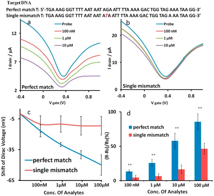
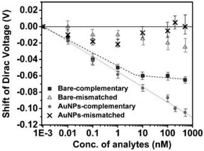
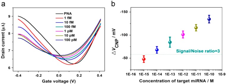
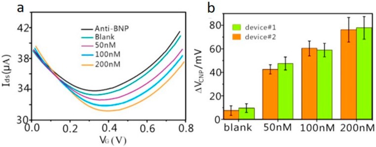
Since the discovery of the two-dimensional (2D) carbon material, graphene, just over a decade ago, the development of graphene-based field effect transistors (G-FETs) has become a widely researched area, particularly for use in point-of-care biomedical applications. G-FETs are particularly attractive as next generation bioelectronics due to their mass-scalability and low cost of the technology's manufacture. Furthermore, G-FETs offer the potential to complete label-free, rapid, and highly sensitive analysis coupled with a high sample throughput. These properties, coupled with the potential for integration into portable instrumentation, contribute to G-FETs' suitability for point-of-care diagnostics. This review focuses on elucidating the recent developments in the field of G-FET sensors that act on a bioaffinity basis, whereby a binding event between a bioreceptor and the target analyte is transduced into an electrical signal at the G-FET surface. Recognizing and quantifying these target analytes accurately and reliably is essential in diagnosing many diseases, therefore it is vital to design the G-FET with care. Taking into account some limitations of the sensor platform, such as Debye-Hükel screening and device surface area, is fundamental in developing improved bioelectronics for applications in the clinical setting. This review highlights some efforts undertaken in facing these limitations in order to bring G-FET development for biomedical applications forward.

Keywords: DNA; Debye length; Dirac voltage; G-FET (graphene-based field effect transistors); antigen binding fragment; aptamer; point-of-care.


Conflict of interest statement

The authors declare no conflict of interest.

Figures

Figure 1
Figure 1
(a) Schematic representation of the conductance band (CB) and valence band (VB) meeting at the K and K’ points of the Brillouin zone at the Fermi level. (b) A schematic representation of a Dirac cone showing in more detail the intersection of the VB and CB at the Fermi level. Adapted from [23]. Copyright 2011 by the American Physical Society.
Figure 2
Figure 2
A schematic representation of the process flow for developing a G-FET for nucleic acid detection. Gold—Contact pads, dark grey—SiO2, light grey—graphene, purple—surface functionalisation.
Figure 3
Figure 3
(a) I-Vg characteristics of a G-FET proposed by Ping et al., highlighting the change in electronic characteristics at each stage of the functionalization and detection process. (b) Dirac voltage shift of increasing concentrations of DNA oligomers of different lengths fitted using the Sips model. Adapted from [25]. Copyright 2016 by the American Chemical Society.
Figure 4
Figure 4
Transfer curves for a G-FET produced by Hwang et al. for each stage of the strand displacement sensing process for (a) perfect match DNA and (b) single nucleotide polymorphism (SNP) DNA. (c) VD shift for perfect match and SNP DNA at various concentrations showing the clear discrimination between perfectly matched DNA and DNA containing an SNP. (d) Quantitative measurement of both the perfectly matched DNA and DNA containing an SNP using resistance changes across the graphene channel. For the data highlighted here ** p < 0.01 based on three sets of independent data points. Reprinted from [38].
Figure 5
Figure 5
Shift in VD for the bare electrode and AuNP decorated G-FET when adding increasing concentrations of complementary DNA and one-base mismatched DNA. Reprinted from [54]. Copyright 2010 by John Wiley and Sons.
Figure 6
Figure 6
(a) Dirac (Vg–IDS) curves for a single G-FET device before and after 100 fM miRNA target exposure. A total of five forward and reverse sweeps were performed on a single device. (b) VD values for each sweep calculated from the graphs depicted in (a). (c) ∆VD values noted for all eight G-FETs on exposure to the target DNA sequence and to the control DNA sequence. It can be noted that the response seen for the control DNA is considerably less than that caused by the target DNA. For the data highlighted here: * p < 0.05; ** p < 0.01, not significant. Adapted by permission from Macmillan Publishers Ltd.: [45], copyright 2014.
Figure 7
Figure 7
(a) The transfer curve of a G-FET decorated with AuNPs with immobilized PNA probe when exposed to increasing concentrations of Let7b miRNA. (b) It can be noted that VD progressively decreases in Vg due to an n-doping effect of the graphene by miRNA hybridization. Reprinted from [41], Copyright 2015, with permission from Elsevier
Figure 8
Figure 8
A schematic representation of the process flow for developing an immuno-based G-FET. Gold—Contact pads, dark grey—SiO2, light grey—graphene, purple—surface functionalisation.
Figure 9
Figure 9
(a) Transfer curves of a PtNPs-decorated rGO-FET in response to brain natriuretic peptide (BNP) in whole blood samples which have been treated with a microfilter; (b) Dirac voltage shift in response to the differing concentrations of BNP. Adapted from [46], Copyright 2017, with permission from Elsevier.
Figure 10
Figure 10
(a) Drain-source current response at the time-dependent introduction of various carcinoembryonic antigen (CEA) concentrations; (b) Drain-source current against CEA concentration fitted based on Hill adsorption model. Adapted from [47], Copyright 2017, with permission from Elsevier.
Figure 11
Figure 11
An illustration highlighting how different ionic buffer solution concentrations affect the screening length (λD). Green—sensor platform, purple—bioreceptor, pink—antigen. Reprinted from [17], Copyright 2011, with permission from Elsevier.
Figure 12
Figure 12
Transfer curves of a G-FET with increasing concentrations (depicted by the arrows) of protective antigen (PA) using an (a) aptamer and (b) antibody. These were then depicted as (c) Dirac voltage shift and (d) change in drain-source current. Reprinted from [48], Copyright 2013, with permission from John Wiley and Sons.

References

    1. Novoselov K.S., Geim A.K., Morozov S.V., Jiang D., Zhang Y., Dubonos S.V., Grigorieva I.V., Firsov A.A. Electric field effect in atomically thin carbon films. Science. 2004;306:666–669. doi: 10.1126/science.1102896. - DOI - PubMed
    1. Kuila T., Bose S., Khanra P., Mishra A.K., Kim N.H., Lee J.H. Recent advances in graphene-based biosensors. Biosens. Bioelectron. 2011;26:4637–4648. doi: 10.1016/j.bios.2011.05.039. - DOI - PubMed
    1. Ali Tahir A., Ullah H., Sudhagar P., Asri Mat Teridi M., Devadoss A., Sundaram S. The application of graphene and its derivatives to energy conversion, storage, and environmental and biosensing devices. Chem. Rec. 2016;16:1591–1634. doi: 10.1002/tcr.201500279. - DOI - PubMed
    1. Clark L.C., Jr., Lyons C. Electrode systems for continuous monitoring in cardiovascular surgery. Ann. N. Y. Acad. Sci. 1962;102:29–45. doi: 10.1111/j.1749-6632.1962.tb13623.x. - DOI - PubMed
    1. Wang J. Glucose biosensors: 40 years of advances and challenges. Sens. Update. 2002;10:107–119. doi: 10.1002/1616-8984(200201)10:1<107::AID-SEUP107>3.0.CO;2-Q. - DOI